What’s the diagnosis?
Recurrent, painful nodules on a girl’s legs


Case presentation
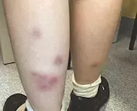
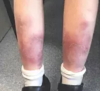
A 10-year-old girl presents with a 12-week history of recurrent, painful, red nodules on her lower legs (Figures 1a and b). Each lesion lasts three to four weeks, resolving with bruising. She has been previously well, with no significant medical or family history.
Differential diagnoses
Conditions to consider among the differential diagnoses of painful, erythematous nodules involving the distal lower limbs in children include the following.
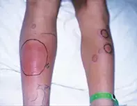
- Cellulitis. In the immunocompetent paediatric population, cellulitis is most commonly caused by Streptococcus pyogenes.1 In contrast to adults, lower limb cellulitis is uncommon in children and the head and neck area more commonly affected; even in adults, bilateral extremity cellulitis is rare.1 Cellulitic areas are swollen, warm and painful, with spreading, well-defined, erythematous borders (Figure 2). There is usually accompanying fever, malaise and chills. Bacterial swabs can be of use to isolate the organism; however, skin swabs are often negative unless there is surface exudate. Other causative organisms include Staphylococcus aureus (methicillin-sensitive or methicillin-resistant) in immunocompetent hosts and atypical organisms such as Pseudomonas aeruginosa in immunocompromised individuals.1
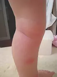
- Insect bite reaction. Insect bite reactions have a variety of morphological appearances but generally appear on exposed areas acutely as grouped, erythematous urticarial papules with associated excoriation due to intense pruritus induced by release of histamine and other immune mediators at the puncture site (Figure 3). Chronic reactions manifest as wheals, urticarial papules and prurigo nodularis-like lesions that can persist for weeks to months after the initial inciting injury and flare with every new insult.2 Exaggerated cutaneous reactions to insect bites can appear as bullous or nodular erythematous lesions, and may indicate underlying infection with Epstein-Barr virus or haematological malignancy (e.g. chronic lymphocytic leukaemia).3
- Panniculitis. The panniculitides represent a diverse group of inflammatory disorders involving the subcutis. All present in a similar way, with painful, poorly demarcated erythematous subcutaneous nodules whose geographic distribution differs with the underlying cause. Classification of the panniculitides is complex, but histologically the conditions are divided on the basis of inflammation (septal, lobular or mixed septal-lobular) and vasculitis (present or absent).4 Causes of panniculitis are generally quite rare, particularly in children, and include infection (Streptococcus spp., Mycobacterium tuberculosis), trauma, cold exposure, alpha-1 antitrypsin deficiency, connective tissue disorders and malignant infiltration of subcutaneous tissue.4
- Bruises. The limbs, particularly the extensor surface of the knees and elbows, pretibial and ‘facial T’ areas are common sites for ecchymoses to occur following accidental trauma in a child.5 However, there are important conditions to exclude in a child with recurrent ecchymoses or those resulting from minimal trauma, such as underlying thrombocytopenias (e.g. thrombotic thrombocytopenic purpura), coagulopathies (von Willebrand disease, haemophilias), vitamin C deficiency, Ehlers-Danlos syndrome, and Henoch-Schönlein purpura.5 It is important to be aware of nonaccidental injury in a child with ecchymoses in areas that are not normally prone to unintentional injury (e.g. ears, neck, cheeks, trunk, proximal extremities and genitalia), associated fractures and inconsistent history, or ‘falls’ in an immobile child.5
- Tuberculid. The term ‘tuberculid’ describes a group of exanthems in individuals with demonstrated immunity to M. tuberculosis as a result of prior infection or sensitisation.6 This includes a condition known as erythema induratum of Bazin, a delayed-type hypersensitivity reaction to tuberculosis antigens, commonly triggered by low temperatures, that manifests as symmetrical subcutaneous nodules with ulceration and subsequent atrophic, hyperpigmented scars that favour the lower limbs.6
- Erythema nodosum (EN). This is the correct diagnosis. EN, which is the most common form of panniculitis at any age, is a hypersensitivity reaction that presents with an acute symmetrical eruption of erythematous, tender subcutaneous nodules and plaques that can arise anywhere but commonly involve the pretibial areas.7 The lesions tend to be poorly defined because they are deep. There may be a history of an upper respiratory tract infection (URTI) or accompanying fever, malaise and arthralgia. In children, a preceding URTI due to Streptococcus spp. is the most common cause, with the URTI preceding the EN nodules by one to three weeks.7 Other causes include infections (e.g. Yersinia, M. tuberculosis), drugs (e.g. oral contraceptive pill), inflammatory bowel disease and sarcoidosis (known as Löffler’s syndrome).7 Up to half of cases are idiopathic. The nodules in EN do not ulcerate and last between two and six weeks, showing spontaneous bruise-like regression without scarring or atrophy (known as erythema contusiformis).7 Chronic EN, which can last for months to years, is characterised by unilateral, migratory nodules that are associated with streptococcal infections and are more common in women.7 Incisional biopsy to the level above the fascia reveals a predominantly septal panniculitis with associated oedema, pathognomonic Miescher microgranulomas (perineutrophilic collections of macrophages) and without primary vasculitic changes.7
Management
The principles of management for EN include symptomatic treatment of the nodules as well as identification of underlying causes, particularly in recurrent or chronic EN lesions lasting longer than six weeks, as seen in this case. Bed rest, leg elevation and analgesia (especially NSAIDs) are first-line symptomatic treatment of EN; however, oral prednisone is effective when there is severe pain and a rapid positive outcome is essential for social reasons. Oral saturated solution of potassium iodide has also been shown to be of some benefit, particularly for chronic EN.7
Outcome
For this child, a biopsy was performed and confirmed the diagnosis of EN. A full blood count and chest x-ray were normal, with only a mild elevation of CRP (9 mg/L). Tests for serum antistreptolysin O titre (ASOT) and angiotensin converting enzyme (ACE) were also negative. As the EN was chronic, the patient was referred for endoscopy and was found to have Crohn’s disease. Subsequent treatment with oral prednisone resolved the skin lesions, and she was referred to a gastroenterologist for follow up.
References
2. Bircher AJ. Systemic immediate allergic reactions to arthropod stings and bites. Dermatology 2005; 210: 119-127.
3. Barzilai A, Shpiro D, Goldberg I, et al. Insect bite-like reaction in patients with hematologic malignant neoplasms. Arch Dermatol 1999; 135: 1503-1507.
4. Requena L, Yus ES. Panniculitis. Part I. Mostly septal panniculitis. J Am Acad Dermatol 2001; 45: 163-183.
5. Kellogg ND; American Academy of Pediatrics Committee on Child Abuse and Neglect. Evaluation of suspected child physical abuse. Pediatrics 2007; 119: 1232-1241.
6. Frankel A, Penrose C, Emer J. Cutaneous tuberculosis: a practical case report and review for the dermatologist. J Clin Aesthet Dermatol 2009; 2: 19-27.
7. Requena L, Yus ES. Erythema nodosum. Dermatol Clin 2008; 26: 425-438.

