What’s the diagnosis?
A woman with pruritic, scaly plaques on her legs

Case presentation
A 31-year-old woman presents with a two-month history of pruritic lesions on her lower legs. She has a history of childhood eczema and asthma, and there is a family history of allergic rhinitis. She has been treated with mupirocin 2% ointment and three courses of oral flucloxacillin 500 mg four times daily, which were ineffective.
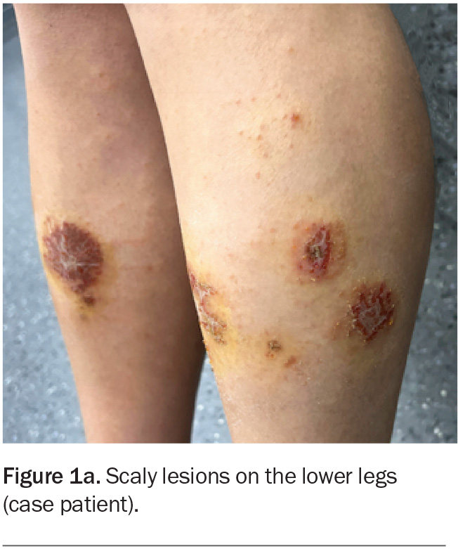
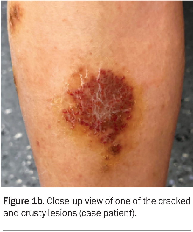
On examination, erythematous, well-circumscribed, scaly plaques, each around 2 to 3 cm in diameter, are observed on the patient’s lower legs bilaterally (Figure 1a, Figure 1b). Some lesions are cracked and crusty but there is no frank pus or discharge. Yellow discolouration, caused by application of povidone-iodine 10% solution, is noted on the lesions and around the periphery.
A bacterial swab is taken from one of the lesions, which returns a positive culture result for Staphylococcus aureus.
Differential diagnoses
Conditions to consider among the differential diagnoses include the following.
Tinea corporis
Tinea corporis is a common dermatophyte infection that typically presents as well-demarcated, annular scaly patches or plaques that are mildly erythematous.1,2 Mild pruritus is common. Tinea corporis is also known as ‘ringworm’, a misnomer that describes how the annular lesions often spread centrifugally and form a central clearing with a raised, active, scaly border on the leading edge. When multiple lesions are present, they can coalesce to form polycyclic plaques.
Tinea corporis is usually caused by anthropophilic species of dermatophytes, most commonly Trichophyton rubrum and Trichophyton tonsurans and less often by zoophilic species, such as Microsporum canis.3
If there is any doubt regarding the diagnosis, a potassium hydroxide (KOH) preparation of skin scrapings can be taken for microscopic examination. The diagnosis is confirmed by visualising dermatophyte hyphae, which appear as bright, linear branching structures.4
For the case patient, the lesions are not typical in appearance of tinea corporis as they do not have a central clearing with an active, scaly border on the leading edge.
Plaque psoriasis
Psoriasis is a chronic inflammatory skin disease that affects 1 to 3% of the population and encompasses a diverse range of heterogeneous presentations.5 The most common type is plaque psoriasis, which accounts for up to 90% of cases.6 Patients may have a single lesion or more. It presents as pruritic, well-demarcated, red or ‘salmon pink’ scaly plaques that can be oval or irregular in shape, surmounted by silvery white scales of varying thickness. When these scales are removed, the underlying smooth and glossy red epidermis with small bleeding points (Auspitz’s sign) is usually revealed.7 Plaques are usually symmetrically distributed over the extensor surfaces of the limbs.
Psoriasis has two peak ages of incidence, the first between 16 and 22 years and the second between 57 and 62 years.8 Adult men and women are equally affected.9 Genetics play an important role in the pathophysiology of psoriasis, although environmental factors are also involved – these include excessive alcohol consumption, streptococcal infections, smoking, obesity and cutaneous trauma.7,10
For the case patient, the distribution of the eruption diffusely across the lower legs is not typical of plaque psoriasis, and the extensor surfaces are spared. The typical silvery scale is also not observed.
Pityriasis rosea
Pityriasis rosea is a relatively common acute self-limiting disease that mainly affects children and young adults. The aetiology is still unknown, but most speculation has centred on viral causes, in particular human herpesvirus 6 and 7.11
The first manifestation of pityriasis rosea is usually the appearance of a herald patch, which is a sharply-defined, erythematous, round or oval plaque, 2 to 5 cm in size with a peripheral collarette of scale. A few days to weeks later, a secondary rash appears: sharply demarcated, pink, oval plaques or patches covered by fine scale. The scale often lags behind the leading edge, which is known as the ‘trailing’ or ‘hanging curtain’ sign.12 This rash typically affects the trunk, upper arms and upper thighs and is often described as forming a ‘Christmas tree-like’ pattern on the upper chest and back because it follows the relaxed skin tension lines. Occasionally, systemic symptoms, such as low-grade fever, malaise and lymphadenopathy, are present.13
Pityriasis rosea is a self-limiting condition. The skin lesions usually fade after three to six weeks and do not require treatment.
For the case patient, the disease course and the distribution of the lesions are not consistent with a diagnosis of pityriasis rosea.
Discoid eczema
This is the correct diagnosis. Discoid eczema, also known as nummular eczema or dermatitis, is a chronic disease with a prevalence of around 2 per 1000 people in the general population.14 It most commonly affects women in early to mid-adulthood. There have been some reports of an increased incidence of atopy in patients affected with discoid eczema.15 Other associations include a history of allergic contact dermatitis, dry skin and excessive alcohol intake.16-18
Discoid eczema is a clinical diagnosis characterised by disc-shaped, circular or oval plaques, mostly 1 to 3 cm in size, that are sharply demarcated, scaly and very pruritic. There are two clinical forms of the disease:
- acute exudative discoid eczema – dull red, serous exudative or crusted plaques
- dry discoid eczema – dry, scaly plaques often with a central clearing.
As the plaques are intensely itchy, they are prone to bacterial infection after excoriation from repeated scratching. If the plaques are crusted or discharging, a bacterial swab should be taken for culture and susceptibility testing. Reports have demonstrated that colonisation of lesions with S. aureus increases severity of the disease.19
For the case patient, the distribution and crusted appearance of the lesions are consistent with a diagnosis of acute exudative discoid eczema. There is a secondary S. aureus infection. In addition, the application of povidone-iodine may have been causing an allergic or irritant contact dermatitis.
Management
The management of discoid eczema (and all other forms of dermatitis) includes general measures, such as avoiding irritants, protecting the skin from injury and applying emollients regularly. These measures are vital and should not be underestimated. Antihistamines may be given for symptomatic relief of pruritus but will not clear the eruption.
A short lukewarm bath, followed by application of a bland emollient, can help the skin to better absorb moisture and relieve pruritus. Dilute bleach baths can decrease inflammation and colonisation of bacteria on the skin.20 Adding one cup of table salt to bath water can reduce stinging during bathing and help to remove dead keratin.21 Dilute bleach baths (prepared as 12 mL bleach per 10 litres of water) can be taken daily for the first one to two weeks of initial treatment. Over-showering and long hot baths should be avoided as they can cause skin irritation and exacerbate discoid eczema.
First-line treatment for discoid eczema involves a potent to very potent topical corticosteroid, such as:
- betamethasone dipropionate 0.05% ointment once or twice daily
- betamethasone valerate 0.1% ointment once or twice daily
- mometasone furoate 0.1% ointment once daily.
This can be combined with a topical antiseptic or antibiotic such as mupirocin 2% ointment. If the rash is extensive or the lesions are discharging, blistered or crusted then oral broad-spectrum antibiotics, such as flucloxacillin, cefalexin, erythromycin or clarithromycin, may be added to the treatment regimen.20
If the rash is still present after two weeks, a potent topical corticosteroid can be used in conjunction with a wet dressing.22 This involves applying the topical corticosteroid, then covering the treated skin with a damp wrung-out dressing; tight fitting cotton clothing may also be used. Alternatively, the ‘soak and smear’ technique may be more practical.22 This involves leaving the affected skin undried after a short lukewarm bath and then applying topical corticosteroid; loose clothes are then worn on top and an emollient is applied the following morning. The use of occlusive dressings (waterproof dressings or plastic film wrap) is another technique that can be useful for localised lesions because it increases absorption of topical corticosteroids and promotes skin rehydration.22
Discoid eczema is a chronic condition and treatment can be challenging. If the patient does not respond to a potent topical corticosteroid and antibiotics, referral to a dermatologist is warranted. Treatment options for troublesome discoid eczema include phototherapy, oral immunosuppressants and dupilumab, a subcutaneously injected monoclonal antibody.22,23
Outcome
The case patient was referred to a dermatologist, who diagnosed discoid eczema. She was advised to stop applying the povidone-iodine solution and was treated with betamethasone dipropionate 0.05% ointment twice daily and a complete course of oral cefalexin 500 mg three times daily for seven days. She was advised to have thrice-weekly bleach baths to prevent future Staphylococcus infection. On review 10 days later, her condition had cleared, with residual dark patches caused by postinflammatory hyperpigmentation. Although this eventually fades with time, it can remain for months or more than one year.
COMPETING INTERESTS: None.
References
1. Leung AKC, Lam JM, Leong KF, Hon KL. Tinea corporis: an updated review. Drugs Context 2020; 9: 2020-5-6.
2. Ely JW, Stone MS, Rosenfeld S. Diagnosis and management of tinea infections. Am Fam Physician 2014; 90: 702-710.
3. Gupta AK, Chaudhry M, Elewski B. Tinea corporis, tinea cruris, tinea nigra, and piedra. Dermatol Clin 2003; 21: 395-400.
4. Richardson MD. Diagnosis and pathogenesis of dermatophyte infections. Br J Clin Pract Suppl 1990 Sep; 71: 98-102.
5. Romiti R. Plaque-type psoriasis – chronic plaque, guttate, and erythrodermic phenotypes. In: Psoriasis, 2nd ed. Menter MA, Ryan C (eds). Boca Raton: CRC Press; 2017: 45-54.
6. Lomholt G. Psoriasis: prevalence, spontaneous course, and genetics: a census study on the prevalence of skin diseases on the Faroe Islands. University of Michigan: GEC Gad; 1963.
7. Burden AD, Kirby B. Psoriasis and related disorders. In: Rook’s textbook of dermatology, 9th ed. Griffiths CEM, Barker J, et al (eds). Chichester UK: Wiley Blackwell; 2016.
8. Henseler T, Christophers E. Psoriasis of early and late onset: characterization of two types of psoriasis vulgaris. J Am Acad Dermatol 1985; 13: 450-456.
9. Hägg D, Sundström A, Eriksson M, Schmitt-Egenolf M. Severity of psoriasis differs between men and women: a study of the clinical outcome measure psoriasis area and severity index (PASI) in 5438 Swedish register patients. Ame J Clin Dermatol 2017; 18: 583-590.
10. Jensen P, Skov L. Psoriasis and obesity. Dermatology 2016; 232: 633-639.
11. Kosuge H, Tanaka-Taya K, Miyoshi H, et al. Epidemiological study of human herpesvirus-6 and human herpesvirus-7 in pityriasis rosea. Br J Dermatol 2000; 143: 795-798.
12. Adya KA, Inamadar AC, Palit A. Dermatoses with “collarette of skin”. Indian J Dermatol Venereol Leprol 2019; 85: 116.
13. Urbina F, Das A, Sudy E. Clinical variants of pityriasis rosea. World J Clin Cases 2017; 5: 203-211.
14. Leung AKC, Lam JM, Leong KF, Leung AAM, Wong AHC, Hon KL. Nummular eczema: an updated review. Recent Pat Inflamm Allergy Drug Discov 2020; 14: 146-155.
15. Jiamton S, Tangjaturonrusamee C, Kulthanan K. Clinical features and aggravating factors in nummular eczema in Thais. Asian Pac J Allergy Immunol 2013; 31: 36-42.
16. Bonamonte D, Foti C, Vestita M, Ranieri LD, Angelini G. Nummular eczema and contact allergy: a retrospective study. Dermatitis 2012; 23: 153-157.
17. Ward S. Eczema and dry skin in older people: identification and management. Br J Community Nurs 2005; 10: 453-456.
18. Higgins EM, Vivier AD. Cutaneous disease and alcohol misuse. Br Med Bull 1994; 50: 85-98.
19. Al-Ameer HA. Detection of secondary bacterial contaminants in patients with eczema. Iraqi Journal of Community Medicine 2007; 20(2): 342-348.
20. Barnes TM, Greive KA. Use of bleach baths for the treatment of infected atopic eczema. Australas J Dermatol 2013; 54: 251-258.
21. Wollenberg A, Barbarot S, Bieber T, et al. Consensus‐based European guidelines for treatment of atopic eczema (atopic dermatitis) in adults and children: part I. J Eur Acad Dermatol Venereol 2018; 32: 657-682.
22. Discoid dermatitis [published 2022]. In: Therapeutic Guidelines. Melbourne: Therapeutic Guidelines. Available online at: https://www.tg.org.au (accessed February 2023).
23. Patruno C, Stingeni L, Hansel K, et al. Effectiveness of dupilumab for the treatment of nummular eczema phenotype of atopic dermatitis in adults. Dermatol Ther 2020; 33: e13290.


