What’s the diagnosis?
A woman with skin-coloured periorbital papules


Case presentation
A 50-year-old woman presents with a four-year history of occasionally pruritic lesions to her periorbital skin, axillae and vulva. The lesions are never painful and are not migratory, with no discharge. They respond well to low-potency topical corticosteroids.
The patient is otherwise well. Her only regular medication is for hypertension, which is controlled. She has a previous history of smoking.
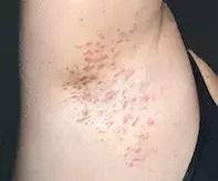
On examination, diffuse skin-coloured papules are observed involving the periorbital and facial skin with symmetrical distribution (Figure 1a). There are also hyperpigmented papules involving the intertriginous skin of the axillae (Figure 1b) and inguinal folds. No abscesses, inflammatory nodules, open comedones, fistulae or sinus tracts are seen.
Differential diagnoses
Conditions to consider in a patient with a symmetrical eruption of skin-coloured papules involving the face and intertriginous skin include the following.
- Hidradenitis suppurativa (HS). This chronic inflammatory disease is characterised by follicular occlusion. HS commonly involves areas located along the ‘milk line’ of apocrine tissue and rarely involves the facial skin. The most common sites of disease (in order of frequency) include the inguinal folds, medial thighs, anogenital skin, chest, axillae and buttocks.1 HS has a global prevalence of 1%.1 Risk factors include female gender (women are affected three times as often as men), metabolic syndrome and a family history of the disease.1 HS is associated with other dermatological conditions, including follicular occlusion syndromes such as acne conglobata, dissecting cellulitis and pilonidal sinus. The primary lesions of HS include painful inflammatory nodules with associated burning, pruritus and hyperhidrosis – these can spontaneously resolve, persist with recurrent inflammatory exacerbations or proceed to an abscess that ruptures. Chronic recurrence of inflammatory nodules and abscesses leads to chronic sinus tracts and fistulae that commonly exude purulent discharge with an offensive odour. The topographic distribution of the vulval and axillary skin is very common, and is seen in 67% and 93% of cases of HS in women, respectively.1 For the case patient, the morphological features of the intertriginous papules are not typical of HS, which are usually painful with a pustular discharge. Furthermore, her facial papules do not appear consistent with acne conglobata that may accompany HS.
- Xanthelasma. Also known as xanthelasma palpebrarum, this is the most common cutaneous form of xanthoma. The incidence increases with age and it is seen usually in individuals who are of middle age and older.2 Onset before the age of 40 years may occur in the context of a lipid disorder such as type II hyperlipoproteinaemia, which has an increased risk of accelerated coronary and peripheral atheroscerosis.3 It is more common in women than men. The lesions of xanthelasma present as yellow-to-pale plaques that are symmetrical and commonly involve the cutaneous surfaces of the inner medial canthi, eyelids and infraorbital skin. These lesions do not involve the axilla, and the involvement of genital and intertriginous areas is not typical. The diagnosis of xanthelasma is made on clinical grounds, based on the location and characteristic appearance of the lesions. Biopsy is rarely required but is diagnostic and demonstrates xanthoma cells (foamy, lipid-laden macrophages) inhabiting the superficial dermis adjacent to blood vessels and adnexal structures.2
- Angiofibromas. These hamartomas appear as multiple small, pink-to-red papules and plaques. They commonly involve the facial skin, particularly the central face (nasolabial folds, cheeks and chin), and are not found on other parts of the body.4 Angiofibromas carry a clinical significance due to an association with systemic diseases such as tuberous sclerosis complex, neurofibromatosis type 2, Birt-Hogg-Dubé syndrome and multiple endocrine neoplasia type 1.5 Biopsy is diagnostic and demonstrates well demarcated, dome-shaped neoplasms consisting of dermal fibroblasts with adjacent dilated blood vessels and collagen fibres.4
- Sebaceous hyperplasia. A common reason for referral to dermatology clinics, sebaceous hyperplasia presents as multiple asymptomatic, small, dome-shaped papules with central umbilication that preferentially occur on the central face (forehead, nose and cheeks). There are, however, reports of sebaceous hyperplasia involving the nipples (Montgomery tubercles), penis (Tyson’s glands) and lips or genitals (Fordyce spots).6 Sebaceous hyperplasia most commonly appears in middle age, although peripubertal onset occurs in familial presenile sebaceous hyperplasia.7 Sebaceous hyperplasia occurs in areas where pilosebaceous units are located, which include almost all body surfaces with the exceptions of the lower lip, dorsal surface of the feet and palmoplantar skin. The dermoscopic features of sebaceous hyperplasia include white-yellow papules with surrounding nonarborising ‘crown’ vessels and milia-like cysts. Biopsy is diagnostic and demonstrates hyperplastic, well differentiated sebaceous glands with no cellular atypia.7
- Syringomas. This is the correct diagnosis. Syringomas are uncommon benign adnexal tumours arising from eccrine and apocrine ducts. They present as multiple small, skin-coloured, pink, yellow or hyperpigmented papules that range between 1 and 3mm in diameter and are symmetrically distributed.8 The most common site of involvement is the periorbital skin, but the scalp, axillae and genital skin can also be affected. There are four clinical variants of syringoma: localised, familial, Down syndrome-associated, and generalised (multiple and eruptive).8,9 Syringomas usually present in early adulthood and are more commonly seen in women. Biopsy is diagnostic and demonstrates groups of small ducts, cysts and epithelial cords in the superficial dermis; the characteristic shape of the cystic eccrine ducts gives rise to the comma-shaped or ‘tadpole’ tail of these neoplasms.8 Although syringomas of the face are usually asymptomatic, when they occur on the vulva they can be very itchy.
Management
Syringomas are benign neoplasms. Management involves excluding associated systemic diseases such as Down syndrome, diabetes mellitus, and Nicolau-Balus, Marfan and Ehlers-Danlos syndromes, as well as improving cosmesis.8,9 Evidence for treatment superiority is lacking and the choice should be tailored to the distribution of the lesions and patient preference. Interventional treatment modalities include carbon dioxide laser, electrocoagulation, intralesional electrodessication, dermabrasion and surgical excision. For lesions located on the vulva, pruritus can be managed with topical corticosteroids, and a skinning vulvectomy is sometimes required for disruptive itching that it refractory to topical therapy.10 Medical management options include topical and oral retinoids.8,9
Outcome
In this patient, a biopsy was performed and confirmed a diagnosis of syringomas. The topographical distribution of her lesions (axillary, genital and periorbital skin) was consistent with the generalised (eruptive) variant. Additional serological tests were performed but did not reveal underlying diabetes mellitus. There were no clinical concerns for connective tissue disease. The patient was referred to a cosmetic dermatologist to discuss treatment options for the facial syringomas, and she opted not to treat her axillary lesions. However, she did continue using mild-potency topical corticosteroids to treat her genital lesions when they were itchy.
References
1. Revuz J. Hidradenitis suppurativa. J Eur Acad Dermatol Venereol 2009; 23: 985-998.
2. Bergman R. The pathogenesis and clinical significance of xanthelasma palpebrarum. J Am Acad Dermatol 1994; 30(2 Pt 1): 236-242.
3. Rohrich RJ, Janis JE, Pownell PH. Xanthelasma palpebrarum: a review and current management principles. Plast Reconstr Surg 2002; 110: 1310-1314.
4. Vincent A, Farley M, Chan E, James WD. Birt-Hogg-Dubé syndrome: a review of the literature and the differential diagnosis of firm facial papules. J Am Acad Dermatol 2003; 49: 698-705.
5. Salido-Vallejo R, Garnacho-Saucedo G, Moreno-Giménez JC. Current options for the treatment of facial angiofibromas. Actas Dermosifiliogr 2014; 105: 558-568.
6. Hogan D, Mohammad S. Sebaceous hyperplasia. Expert Review of Dermatology 2011; 6(1): 91-96.
7. Wang Q, Liu JM, Zhang YZ. Premature sebaceous hyperplasia in an adolescent boy. Pediatr Dermatol 2011; 28: 198-200.
8. Williams K, Shinkai K. Evaluation and management of the patient with multiple syringomas: a systematic review of the literature. J Am Acad Dermatol 2016; 74: 1234-40e9.
9. Soler-Carrillo J, Estrach T, Mascaro JM. Eruptive syringoma: 27 new cases and review of the literature. J Eur Acad Dermatol Venereol 2001; 15: 242-246.
10. Rettenmaier MA, Braly PS, Roberts WS, Berman ML, Disaia PJ. Treatment of cutaneous vulvar lesions with skinning vulvectomy. J Reprod Med 1985; 30: 478-480.
Skin lesions

