The floppy baby: a diagnostic challenge
Neuromuscular disorders
Infant and newborn development
The prognosis and treatment of infantile hypotonia are contingent on the underlying diagnosis. Distinguishing between normal and abnormal hypotonia, and central and peripheral causes, is key in initiating a diagnostic pathway for affected babies.
- In most infants with hypotonia the condition is benign and resolves spontaneously; however, in a small number it is a symptom of a serious underlying neurological disorder.
- The GP is the first port of call for parents concerned by floppiness, low muscle tone or weakness in their child, and can initiate the diagnostic pathway for infants affected by hypotonia.
- The primary question on examining the floppy baby is whether they are floppy and strong, which suggests a CNS disorder, or floppy and weak, suggesting a peripheral nervous system problem.
- Most babies require only a few screening tests for hypotonia, but early referral to a paediatrician or paediatric neurologist should be considered if the hypotonia is likely to have a central or peripheral cause.
- Early treatment with the novel drug nusinersen has recently been shown to improve motor development and increase life expectancy in babies with spinal muscular atrophy types 1 and 2.
The floppy infant is a diagnostic challenge to the general practitioner. Hypotonia, which reflects decreased muscle tone and diminished resistance to passive movement, is the most common presenting sign in newborns and young babies with central or peripheral nervous system disorders. In most infants with the condition, hypotonia is benign, resolving spontaneously in the second or third year of life; however, in a small number, it is a symptom of a serious underlying neurological disorder. Distinction between benign and pathological hypotonia is contingent on a careful assessment and, sometimes, repeated reviews to determine the baby’s developmental trajectory. As with other assessments, the evaluation should begin with a targeted history and physical examination.
Identification of the cause of infantile hypotonia is important to enable a definitive diagnosis and specific treatment. Low muscle tone can be caused by a variety of conditions (Table 1). Hypotonia is usually central (i.e. caused by disorders of the brain or spinal cord). Only 15 to 30% of cases are caused by peripheral abnormalities, which may occur at any neurological level between the spinal cord and motor unit (Figure 1). Such disorders of the anterior horn cell, peripheral nerve, neuromuscular junction or muscle are nevertheless very important to recognise, as new therapies with the potential to markedly improve outcomes for affected children have recently been identified.
Medical history
The pregnancy and birth history may provide clues to a possible asphyxial insult, which may have predisposed the baby to hypoxic–ischaemic encephalopathy (HIE). HIE is a very common cause of neonatal hypotonia, but should evolve over the first few months of life to a picture of increasing tone and early spasticity, with delayed development of motor and other milestones. A history of seizures or hypoglycaemia may also point to central causes of hypotonia.
Examination findings
Floppy babies can be strong or weak. Weak infants always have hypotonia, but hypotonia may exist without weakness. The primary question on examining the floppy baby is whether they are floppy and strong, which suggests a central problem, or floppy and weak, suggesting a peripheral condition (Table 2). Examination of muscle bulk, power and tendon reflexes and for the presence or absence of ptosis, ophthalmoplegia and tongue fasciculations can largely define the anatomical site of the neurological problem.
Infants with central hypotonia have low tone but have volitional antigravity movement of the extremities and their deep tendon reflexes are often increased, sometimes with overt spasticity and clonus. Facial movement is generally normal, but these babies may appear dull and less responsive than usual. Infants with Down syndrome and other genetic or chromosomal conditions may have characteristic dysmorphic features and are often delayed in all domains, not just in their motor development.
Infants with peripheral causes of hypotonia have decreased spontaneous movement, absent antigravity movement of the proximal arms and legs and decreased or absent deep tendon reflexes. Intelligence and facial movement may be normal or the baby may have ptosis and/or weakness of the extraocular muscles. Anterior horn cell disorders such as spinal muscular atrophy are associated with tongue fasciculations. Peripheral causes of hypotonia are sometimes associated with congenital or postnatally developing joint contractures.
Children with hypotonia but otherwise normal neurological development and physical examination most likely have benign congenital hypotonia and can be monitored clinically, without undergoing further investigations and treatment.
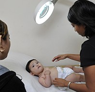
Decreased tone is demonstrated by the resting position when supine: the floppy baby lies in a ‘frog leg’ position with the hips abducted and externally rotated. There is marked head lag on a ‘pull to sit’, and the baby hangs in an inverted-U position when held in horizontal suspension rather than extending the neck against gravity (Figures 2a and b).
What tests should be ordered in floppy babies?
Most babies require only a few screening tests for hypotonia, but early referral to a paediatrician or paediatric neurologist should be considered if the hypotonia is likely to have a central or peripheral cause.
Chromosomal testing should be considered in children with hypotonia and dysmorphism. Hypothyroidism is a rare but treatable cause of low tone and developmental delay that should also be considered. The serum creatine kinase level, which reflects muscle injury, may be increased for a few days after birth but should then normalise. Persistent elevation after one week of age should prompt an evaluation for a muscle disease.
Cranial ultrasonography is widely available and has the advantages that it does not require sedation and carries no radiation exposure. More advanced neurological investigations and imaging should be undertaken under guidance from a paediatrician or paediatric neurologist.
Outcomes of hypotonia
The outcome of infantile hypotonia depends on the underlying cause. Prognosis and treatment are therefore contingent on the specific diagnosis.
Typically, benign hypotonia will improve over time as the child grows. However, an infant who is initially hypotonic due to hypoxic–ischaemic brain injury will usually develop increasing spasticity, with static cognitive impairment, and ultimately will be diagnosed with cerebral palsy.
The natural history of hypotonia in genetic disorders is variable. Children with Down syndrome and other chromosomal conditions remain hypotonic, while tone tends to normalise with age in children with Prader–Willi syndrome and other similar conditions.
Weakness has a different prognosis depending on the specific diagnosis. In some cases, such as spinal muscular atrophy presenting in early infancy, weakness can be rapidly progressive and life limiting. In congenital myopathies and muscular dystrophies, weakness is usually slowly progressive into adulthood.
Spinal muscular atrophy
The most severe form of peripheral hypotonia in infants is caused by spinal muscular atrophy (SMA). SMA is the paediatric equivalent of adult motor neuron disease. It affects the motor neurons of the spinal cord, resulting in muscle weakness and wasting. Life expectancy can be decreased in children with SMA; in the most severe cases it is less than two years.
SMA is a genetic disorder caused by recessive mutations in the survival motor neuron 1 (SMN1) gene. When the SMN1 gene has reduced function, motor neurons in the spinal cord and brainstem die prematurely. In babies with SMA, this process starts before birth, so some children are symptomatic in the first few weeks or months of life.
SMA has several forms that present at different ages and have variable severity. The most common and severe form, SMA type 1 (previously known as Werdnig– Hoffman disease) presents before six months of age. Typical features of SMA type 1 include:
- severe weakness with low muscle tone
- poor head control
- weak cry
- weak cough
- difficulty with swallowing and handling of oral secretions
- motor delay and an inability to sit.
Babies with SMA develop a bell-shaped chest from using accessory muscles in breathing. They are also increasingly prone to severe respiratory tract infections and aspiration pneumonia.
The process of reaching a diagnosis of SMA generally begins when parents, a maternal and child health nurse or the GP raise concerns about floppiness, low muscle tone or weakness in a baby or small child. Because these symptoms can be benign and can relate to many other conditions, they are often dismissed. The GP can be the health professional who initiates the diagnostic pathway for affected children, and should prospectively monitor the child’s condition.
On suspicion of this condition, the GP should refer the infant urgently to a paediatric neurologist with expertise in diagnosis and management of paediatric neuromuscular disorders (Box). A blood test can confirm whether an infant has the SMN1 mutation causing SMA. Early recognition and referral could have a significant long-term effect on the lives of babies affected by SMA.
New developments
Until recently, SMA has been managed similarly to other hypotonic conditions. Treatment has been symptomatic, based on comfort measures and management of complications of weakness and of feeding and breathing difficulties. Treatment in a multidisciplinary paediatric neuromuscular clinic with involvement of a neurologist, respiratory specialist, genetic counsellor, physiotherapist, occupational therapist and dietitian decreases hospitalisations and prolongs life.1,2 In this setting, the GP, as first point of call for managing intercurrent illnesses, has had a crucial role in facilitating rapid treatment of chest infections or feeding difficulties.
This approach has now been altered radically with the identification of a novel therapeutic agent, nusinersen, which contains an antisense oligonucleotide, a molecule that works to compensate for the reduced function of the SMN1 gene. Early treatment with nusinersen has recently been shown to improve motor development and increase life expectancy in babies with SMA types 1 and 2.
In Australia, nusinersen has now been approved by the TGA but has not yet been approved by the Pharmaceutical Benefits Advisory Committee for subsidisation. It has been approved for treatment of all forms of SMA in the United States, where it costs around AU$500,000 per patient per year. This cost raises challenging ethical questions regarding the funding of ultraexpensive drugs for rare diseases.
Conclusion
In general practice, most babies presenting with low muscle tone will not have an underlying neurological problem. For the small number in whom testing and monitoring raise suspicion of a central or peripheral abnormality, early referral by the GP to a paediatrician, paediatric neurologist or neuromuscular clinic, as appropriate, is crucial to achieving the best possible outcome for each infant. MT
References
spinal muscular atrophy. Part 2: Pulmonary and acute care; medications, supplements and immunizations; other organ systems; and ethics. Neuromusc Dis 2018; 28: 197-207.