
Sessile pigmented lesions
Skin conditions
Case presentation
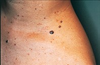
Over a 10-year period, a 52-year-old woman developed a series of pigmented papular lesions on her neck and upper trunk (Figure 1). Dermoscopic examination of the largest lesion showed a well demarcated symmetrical lesion with a smoky tan to light brown colour and numerous yellowish-white globules (Figure 2). The surrounding small papular lesions showed identical findings. A shave biopsy revealed a hypertrophic epidermis associated with small keratinocytes that enclosed multiple large keratin-filled pseudocysts (Figure 3).
Single article purchases are temporarily unavailable due to site maintenance.
If you would like to purchase an article during this time, please email us at [email protected] with the article details and we'll assist you directly. We'll also let you know when online purchasing is available again.
Thank you for your patience and understanding.