A friable pigmented lesion
Skin lesions
Skin conditions
Case presentation
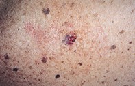
An 82-year-old man with multiple seborrhoeic keratoses developed an irritable pigmented lesion (measuring 1.3 cm x 1 cm) on his back (Figure 1). This had bled intermittently over a six-week period and had been itchy. Dermoscopy revealed an irregular pigmented lesion that had numerous blue–black dots giving a stippled pattern with pale scar-like areas adjacent to a raw bleeding surface (Figure 2). Excision biopsy showed an eroded epidermis with haemorrhage. The upper dermis contained abundant melanin pigment, lymphocytic inflammation and fibrosis. The adjacent epidermis was papillomatous and contained isolated keratin pseudocysts, but there was no melanocytic proliferation or atypia (Figure 3).
Single article purchases are temporarily unavailable due to site maintenance.
If you would like to purchase an article during this time, please email us at [email protected] with the article details and we'll assist you directly. We'll also let you know when online purchasing is available again.
Thank you for your patience and understanding.