Seborrhoeic keratosis with a black nodule
Skin lesions
Skin conditions
Case presentation
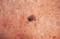
A 56-year-old man presented with a smooth shiny black nodule (1.8 cm in diameter) that was superimposed on a longstanding seborrhoeic keratosis on his chest wall (Figure 1). The nodule had appeared over a six-month period between skin checks. Dermoscopy revealed a central homogeneous jet black surface and irregular periphery that merged with a blue–grey honeycombed corona containing linear and small round sandy deposits. The outer margin had an irregular border with further dark centres that merged with mottled sun-damaged skin (Figure 2). Excision biopsy showed a seborrhoeic keratosis with a hyperplastic epidermis, hyperpigmentation, keratin pseudocysts and inflammation, but no atypia (Figure 3).
Single article purchases are temporarily unavailable due to site maintenance.
If you would like to purchase an article during this time, please email us at [email protected] with the article details and we'll assist you directly. We'll also let you know when online purchasing is available again.
Thank you for your patience and understanding.