Peer Reviewed
Perspectives on dermoscopy
A solitary pigmented nodule on the thigh
Recent articles on:
Skin lesions
Skin lesions
Abstract
This series will help clinicians with an interest in dermoscopy. This month, we present a 55-year-old woman with an asymptomatic nodule on her thigh.
Key Points
- With sufficient training and expertise, clinicians can use dermoscopy to improve diagnostic accuracy for melanocytic lesions and other common skin tumours.
Case presentation
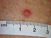
A 55-year-old woman presented with an asymptomatic pigmented nodule on her left thigh that had been present for six months (Figure 1). The lesion was approximately 6 mm in maximal diameter and firm on palpation. Lateral compression of the lesion resulted in dimpling of the nodule. Dermoscopy revealed a central, white, scar-like patch with a delicate peripheral pigment network in association with vertically orientated telangiectatic vessels (Figure 2).
Get full access
Buy this article
Single article purchases are temporarily unavailable due to site maintenance.
If you would like to purchase an article during this time, please email us at [email protected] with the article details and we'll assist you directly. We'll also let you know when online purchasing is available again.
Thank you for your patience and understanding.
Already a subscriber? Login here.