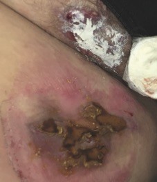
Chronic inflammatory nodules in the groin area
Test your diagnostic skills in our regular dermatology quiz. What are the chronic inflammatory nodules affecting this woman’s groin area?
Case presentation
A 36-year-old woman presents with a longstanding history of chronic inflammatory nodules in the groin area (Figure 1). The lesions first appeared around puberty and have never fully resolved. They are extremely painful and she has had to take time off work in the past because of the pain.
Skin swabs have been performed previously but have not returned any organisms. The patient has tried multiple short courses of cephalexin over the past decade and washes regularly with triclosan 1% lotion and bleach baths, but there has been no improvement. Her father has had similar lesions, mainly affecting his axillae and apron area.
The patient is overweight and she has been diagnosed with polycystic ovary syndrome. She smokes 10 cigarettes per day.
On examination, multiple tender subcutaneous nodules are visible in the groin area with surrounding erythema. There is also a draining sinus with serous ooze; dressings that have been on the groin area are stained with this exudate. The patient has scarring and contractures in the axillae from previous surgical drainage of similar inflammatory nodules.
Differential diagnoses
Conditions to consider among the differential diagnoses include the following.
- Folliculitis and carbuncles. These conditions are caused by Staphylococcus aureus. They are unlikely to be the correct diagnosis for this patient, for whom previous skin swabs have been negative and treatment with antibiotics has been unsuccessful.
- Crohn’s disease. This chronic inflammatory disease can affect any part of the gastrointestinal tract. Perianal characteristics include metastatic granulomatous ulcers and nodules and anal fissures or fistulae, which can precede symptoms of intestinal disease. If Crohn’s disease is suspected then a faecal calprotectin study and endoscopic visualisation, with or without biopsy of the gastrointestinal tract, should be performed. This patient did not have perianal ulcers, anal fissures or fistulae.
- Granuloma inguinale. This atypical infection might be considered in this case, given the failure of previous short courses of antibiotics. Granuloma inguinale is a genital ulcerative condition that has largely been eradicated in Australia but cases still occur in Papua New Guinea, South Africa and Brazil. The most common lesions of granuloma inguinale are friable, beefy red ulcers that start as papules or subcutaneous nodules and are typically not painful. Skin biopsy is required for a diagnosis of granuloma inguinale to identify characteristic Donovan bodies. It is unlikely to be the diagnosis in the case of this patient, whose lesions are very painful.
- Hidradenitis suppurativa. This is the correct diagnosis. Hidradenitis suppurativa (HS) is an inflammatory skin disease that follows a painful, chronic course. Onset is usually after puberty. It is a disease of the hair follicle that results in painful, deep-seated nodules and abscesses that rupture and lead to sinus tracts and scarring. HS occurs in areas of skinfolds – axillae, anogenital and infra-mammary areas (Figure 2 and Figure 3). It has a significant impact on a patient’s quality of life and there is often a long delay to diagnosis. There is an association with comorbid diseases such as pyoderma, inflammatory arthritis, obesity, inflammatory bowel disease, polycystic ovary syndrome and metabolic syndrome.
Management
HS is a clinical diagnosis. The investigations selected for an individual patient will be determined by the treating physician on the basis of the history and examination.
For mild to moderate cases of HS (i.e. an absence of deep inflammatory lesions), topical clindamycin lotion 1 to 2% can be used, twice daily for three months.
If topical therapy is unsuccessful, a number of systemic therapies may be tried. First-line treatment is long-term oral tetracyclines, doxycycline and minocycline. HS appears in many cases to be androgen-dependent, and antiandrogen treatment is often successful. This is most easily achieved with spironolactone 100 to 200mg daily;1 however, the use of other agents such as cyproterone acetate 2.5 to 5mg daily and finasteride 5mg daily has been described.2 The antibiotic combination of clindamycin (300mg twice daily) and rifampicin (either 600mg once daily or 300mg twice daily) can be effective, particularly in moderate to severe disease. The tumour necrosis factor (TNF) alfa antagonist adalimumab (40mg subcutaneous injection weekly) was approved in 2016 as second-line treatment for moderate to severe HS.3 There are anecdotal reports of other TNF-alfa blocking agents being effective (off-label use).
A retrospective database review has shown that early-onset HS is associated with stronger genetic susceptibility, more widespread disease and endocrine comorbidities.4 Therefore, metformin (up to 1500mg) may be useful for patients with early-onset HS.
Surgical therapy is often necessary for patients with HS and should be decided on a case-by-case basis, depending on the type and extent of scarring. Surgical options include deroofing procedures, laser treatment, and local or wide excision. There are case reports of squamous cell carcinoma (SCC) complicating HS, with human papillomavirus infection and longstanding anogenital disease implicated as contributing factors, particularly in men.5 Therefore, suppurating, ulcerative or irregular masses in the anogenital area particularly should be biopsied to rule out SCC. A biopsy should be performed if there is any suspicion of malignancy.
Adjuvant therapies for HS include pain management, treatment of superinfection, laser hair removal and education regarding appropriate dressings. Patient counselling about weight loss and smoking cessation may also be appropriate. Useful patient information about HS management is also available online (www.hs-online.com.au). HS has the potential to scar and has a large impact on quality of life. Early referral of any patient with recurrent groin nodules where swabs have not returned S. aureus can prevent many years of distress.
Outcome
The case patient was treated with spironolactone 100mg daily, which was well tolerated. The associated irritant dermatitis, which arose from chronic use of talcum powder and iodine under occlusion of scented make-up pads as dressings, was treated with 1% hydrocortisone ointment, emollient and a soap substitute. When the irritant reaction had settled, spironolactone was ceased and the patient commenced treatment with 2% clindamycin cream, which she applied daily after showering. She was encouraged to stop smoking and to embark on a sensible eating plan.
Over the next three months, there was slow resolution of the skin eruption. The patient has commenced treatment with adalimumab, 40mg subcutaneous injection weekly, and has experienced a significant reduction in inflammatory nodules over a three-month period. MT

