
July 2023
Ultrasound assessment of paediatric forearm fractures may reduce need for x-rays
By Rebecca Jenkins
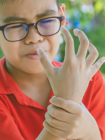
Children with a suspected distal forearm fracture have no-worse arm function at four weeks if initially assessed with ultrasound rather than with x-ray, an Australian study shows.
In an open-label, non-inferiority trial at four centres in southeast Queensland, researchers enrolled 270 participants aged 5 to 15 years of age who presented to the emergency department with an isolated distal forearm injury, without a clinically visible deformity, in whom imaging was indicated.
Participants were randomly assigned to point-of-care ultrasonography or radiography and then followed for eight weeks, with the primary outcome physical function of the affected arm at four weeks measured with the Pediatric Upper Extremity Short Patient-Reported Outcomes Measurement Information System (PROMIS).
Of the 262 participants with available data, PROMIS scores in the ultrasonography group were noninferior to those in the radiography group (mean 36.4 and 36.3 points, respectively; mean difference 0.1 points, which was within the five points noninferiority margin).
‘No clinically important fractures were missed and there were no between-group differences in the occurrence of adverse events,’ the study authors wrote in The New England Journal of Medicine.
Lead author, Dr Peter Snelling, Senior Lecturer at the School of Medicine and Dentistry at Griffith University and Emergency Physician at Gold Coast Hospital and Health Service, told Medicine Today they saw a two-thirds reduction in the number of x-rays required when ultrasound was used first, with the patients who did not require further radiography discharged immediately.
‘It also saved about 15 minutes per patient being in the emergency department and the families actually preferred it over x-ray,’ he said.
Ultrasound had become more portable, affordable and is now available in almost every major emergency department in Australasia, he noted.
Given a variety of clinicians were trained to carry out the ultrasound imaging using different devices, the protocol could also potentially be used in a range of settings, including general practice and the sports field.
‘What about children based in regional and remote settings who have to travel hundreds of kilometres to have an x-ray? This could potentially save that journey,’ Dr Snelling said.
Dr Snelling was keen to develop a self-sustaining training pathway to boost the numbers of people who could train other healthcare professionals to use ultrasonography.
There had also been interest in the protocol in Nigeria, where children were seen in the community and managed without any imaging with poor outcomes from missed fractures.
‘This is an option to potentially improve the lives of children globally,’ Dr Snelling said.