Peer Reviewed
Perspectives on dermoscopy
A new plum-coloured papule
Recent articles on:
Melanoma
Melanoma
Recent articles on:
Skin cancer
Skin cancer
Abstract
Dermoscopy is useful in detecting the blue–black melanin pigment in cutaneous melanoma metastasis, particularly when the lesions may simulate angiomas.
Key Points
Case presentation
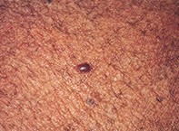
Over a two-month period, a 76-year-old man developed a smooth plum-coloured papule (measuring 7 mm x 4 mm in diameter) on his left thigh (Figure 1). Dermoscopy of the papule revealed an oval grey–blue lesion with a superimposed milky veil (Figure 2). The surrounding skin was sun-damaged and had mottled pigment. The excision biopsy contained a subepidermal nodule consisting of sheets of pleomorphic and focally pigmented melanocytes separated by a vascular network (Figure 3). Review of the patient’s history revealed that nine years before the consultation, a melanoma had been removed from his left calf. Over the ensuing five years, four solitary metastases localised to the left leg had been surgically excised.
Purchase the PDF version of this article
Already a subscriber? Login here.