Peer Reviewed
Perspectives on dermoscopy
A new pigmented lesion
Recent articles on:
Melanoma
Melanoma
Recent articles on:
Skin cancer
Skin cancer
Abstract
Dermoscopy is helpful in evaluating deeply pigmented skin lesions because the multicomponent pattern is highly associated with melanoma.
Key Points
Case presentation
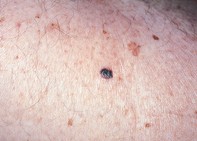
Over a six-month period, a 56-year-old man developed a new pigmented lesion, 0.8 cm in diameter, over his right lateral thigh (Figure 1). Dermoscopy revealed a dark asymmetrical lesion with multiple components and colours. The border was associated with irregular, blunt, pigmented projections (pseudopods). There were irregularly sized pigmented globules and dots that extended to the periphery and a prominent milky veil (Figure 2). Excision biopsy showed large nests of atypical melanocytes within the epidermis and upper dermis. Some of the nests were fused together and were associated with lymphocytic inflammation and pigment release into the surrounding dermis (Figure 3).
Purchase the PDF version of this article
Already a subscriber? Login here.