What’s the diagnosis?
An enlarging lesion on the neck


Case history
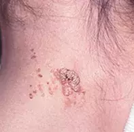
A 13-year-old boy presented with a raised, hyperkeratotic, warty lesion on his neck (Figure 1). His parents said it had been there since his first year of life but had recently enlarged and become more prominent. The patient’s voice broke recently. The lesion is not symptomatic but it often gets caught on his collar.
Diagnosis
The correct diagnosis in this case is a verrucous epidermal naevus, a birthmark that occurs mainly on the trunk and limbs. These lesions may be congenital but in over half of cases the onset is in the first year after birth. They may spread beyond their original size with age, usually over a few months but sometimes several years. It is not unusual for them to become more raised and ‘warty’ at puberty.
The colour of verrucous epidermal naevi ranges from flesh-coloured to brown and black. They may occur as single or multiple grouped lesions and can have a linear or whorled distribution. On the chest there is often a dramatic cut-off at the midline.
Differential diagnosis
Although verrucous epidermal naevi resemble common viral warts (hence the name verrucous) and seborrhoeic keratoses, they have little in common with these lesions.
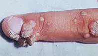
- Viral warts are very unlikely to have their onset in a patient’s first year of life or to persist unchanged for more than two or three years. It is also unusual for many viral warts to be grouped on the shoulder or neck (as in this patient’s case), although this presentation is not uncommon on the hands (Figure 2) and feet.
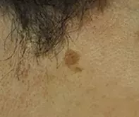
- Seborrhoeic keratoses generally occur from middle age and they present as discrete lesions (Figure 3). The back is a typical location, but they can be found on any part of the skin.
Cause
The cause of verrucous epidermal naevi is unknown, but they are believed to arise from a post-zygotic mutation resulting in epidermal mosaicism.
The lesions are harmless and have no potential for malignant transformation and, particularly when localised, are rarely associated with any other abnormalities. They can easily be differentiated from a viral wart on histopathology, but they may be confused with a seborrhoeic keratosis unless the pathologist is aware of the age of the patient.
Treatment
Treatment is often requested for verrucous epidermal naevi for cosmetic or functional reasons. Although there are superficial ablative procedures such as laser ablation that are effective, these lesions will often recur afterwards. Naevi that have any protruding areas or are interfering with function need complete excision to avoid recurrence.
Skin lesions

