What’s the diagnosis?
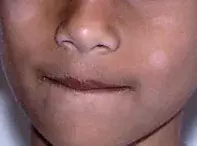
Hypopigmented and scaly lesions on a boy's cheek

Figure. The hypopigmented lesions on the boy’s cheeks.
Case presentation
A 6-year-old boy presents with hypopigmented and slightly scaly lesions on his cheeks that were first noticed two months earlier (Figure). They are not causing symptoms.
Differential diagnosis
Conditions to include in the differential diagnosis in a child of this age include the following.
- Pityriasis versicolor. This chronic, harmless reaction to Malassezia yeast, a cutaneous commensal organism, presents as scaly macules that can be hypopigmented or hyperpigmented. The distribution is patchy and there are often large numbers of macules that can coalesce and produce larger lesions. The condition is unusual in children but when it occurs the lesions are usually on the face (unlike in adults, for whom the lesions are most commonly on the torso). Sometimes they cause a small degree of irritation, but most patients are asymptomatic. Pityriasis versicolor most frequently occurs in hot and humid climates.
- Post-inflammatory hypopigmentation. This usually presents as diffuse loss of pigment (not as complete depigmentation) and can be caused by any inflammatory or traumatic process. The most common causes in children are injury, atopic dermatitis, psoriasis and skin infections. It is much more apparent in children who have darker skin.
- Vitiligo. This autoimmune depigmenting skin disorder results in complete loss of melanocytes in affected skin. The lesions themselves are characteristically very well defined, ivory white in colour and not scaly.
- Halo naevus. A halo naevus occurs when a small pre-existing melanocytic naevus develops a surrounding area of hypopigmented skin. Following formation of the halo, the naevus typically regresses through autoimmune-mediated destruction of melanocytes, leaving behind an area of hypopigmentation without any scale. These lesions are most commonly located on the back. They are not typically as large as the lesions seen in this patient and the presence of multiple simultaneously hypopigmented lesions would be unusual.
- Pityriasis alba. This is the correct diagnosis. Pityriasis alba is a common skin condition limited to children and adolescents. It presents as flat, scaly and hypopigmented patches and sometimes with a small degree of erythema. There is usually more than one lesion, and they are usually small in size and irregular. Pityriasis alba commonly presents on the face, particularly on the cheeks, but it can also affect the limbs and trunk. Lesions do not fluoresce under Wood’s lamp. Children are frequently asymptomatic, but the lesions are often preceded by a nonspecific dermatitis. It is often more difficult to visualise pityriasis alba in children with fair skin, and although it occurs in all skin types it tends to be more obvious in individuals who have darker skin. In the case described above, the child presented at the end of summer, when the lesions were more prominent.
Management
Pityriasis alba is a benign self-limiting condition. The lesions tend to be asymptomatic and most children do not require treatment. Emollient creams, however, can help to reduce scale and mild topical corticosteroids (e.g. 1% hydrocortisone) can settle any residual inflammatory process. After the underlying condition is controlled, sun exposure will encourage repigmentation.
Recent articles on:
Skin lesions
Skin lesions

