What’s the diagnosis?
A red nodule on the finger
Case presentation
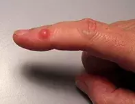
A healthy 54-year-old man presents with a red nodule on his right index finger. The lesion was of slow onset and was not tender, but it was slightly compressible and could be transilluminated.
Diagnosis
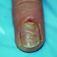
Digital myxoid (mucous) pseudocyst is the correct diagnosis in this case. This is a benign, cystic swelling that occurs on the fingers between the distal interphalangeal joint and the nail bed; it may involve the nail bed and cause a groove on the nail (Figure 2). These lesions are not true cysts, but histopathology reveals a fibrous capsule containing a myxomatous stroma. The clear gelatinous fluid of a myxoid pseudocyst can easily be expressed after piercing the surface with a sterile needle. This simple procedure drains the lesion and confirms the diagnosis.
Aetiology
The fluid in the pseudocyst is thought to originate from the distal interphalangeal joint with which there is usually a communication. Osteoarthritis may be a factor in damaging the joint capsule so that this occurs.
Differential diagnosis
There are several non-tender lesions that occur on the fingers.
- Granuloma annulare is a harmless idiopathic condition with a characteristic granulomatous histopathology. The classic lesion is a slightly raised, erythematous and non-scaly ring, but granuloma annulare can present with discrete papules, particularly on the fingers. The lesions of granuloma annulare are not as raised or as red as the lesion shown in Figure 1.
- Insect bites can occur on exposed skin and are usually very inflammatory. They are of acute onset and invariably painful and itchy.
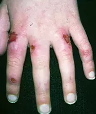
- Chilblains are commonly found on the fingers (Figure 3). Patients who are prone to chilblains have a peripheral circulation that reacts with excessive vasoconstriction to cold conditions; they develop chilblains after exposure to the cold, most often just by going outdoors in cold conditions without warm gloves. Chilblains are of acute onset, itchy and sore, and they erode rapidly to form crusted lesions.
- Warts are very common on the fingers but they are not erythematous. They generally have a rough surface but occasionally may appear smooth, particularly when they are not fully developed. However, warts are never as red as the lesion shown in Figure 1.
- Giant cell tendon sheath tumours occur over the distal interphalangeal crease in patients with osteoarthritis. They are firm and rubbery and cannot be transilluminated.
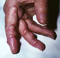
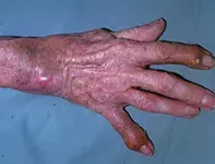
- Rheumatoid nodules present as firm, raised pink lesions that are prone to ulceration (Figure 4). They occur in patients with rheumatoid arthritis.
- Gouty tophi occur in patients with gout in the setting of acute arthropathy (Figure 5). They may ulcerate and exude tophaceous material.
Management
There are many minor destructive procedures that are effective for myxoid pseudocysts. After a lesion is drained, cryotherapy followed by firm bandaging for a week is often successful conservative treatment. Intralesional corticosteroid, laser ablation and sclerotherapy have also been described to treat these lesions. Myxoid pseudocysts treated conservatively often recur, but many patients prefer re-treatment with a conservative approach over surgery, which is definitive. However, a surgical approach is often necessary for a lesion that involves the nail fold because the lesion itself is not accessible for conservative management strategies.
Skin lesions

