
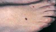
A mole with central dark branches
Melanoma
Case presentation
Over a three-month period, a 45-year-old woman noted progressive darkening of a longstanding mole, measuring 6 mm in diameter, on the dorsum of her right foot (Figure 1). Dermoscopy showed an asymmetrical mole with an irregular pigment network with regional variation in intensity. A darkly pigmented branched component with globules and dots was in the centre of the mole (Figure 2). Excision biopsy showed nests of atypical melanocytes within the epidermis and upper dermis, which were obscured by a marked lymphocytic reaction. The surface of the skin had collections of red cells, melanocytes and melanin pigment between keratin filaments (Figure 3).
Single article purchases are temporarily unavailable due to site maintenance.
If you would like to purchase an article during this time, please email us at [email protected] with the article details and we'll assist you directly. We'll also let you know when online purchasing is available again.
Thank you for your patience and understanding.