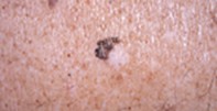
A pigmented lesion with a white patch
Melanoma
Skin lesions
Case presentation
A 75-year-old man with extensive sun-damaged skin presented with an irregularly pigmented lesion of unknown duration, measuring 0.8 cm x 1.0 cm, on his back. The pigmented portion formed a crescent around a white patch (Figure 1). Dermoscopy revealed an irregularly pigmented lesion with a coarse and broken pigment network and streams of pigment at the edge. There were multiple blue–black dots and scattered pale areas within the pigmented segment. The large white patch appeared structureless (Figure 2). The excision specimen showed an atrophic epidermis with confluent atypical melanocytes present along the junctional zone together with extensive dermal fibrosis containing clumps of melanin pigment and lymphocytes (Figure 3).
Single article purchases are temporarily unavailable due to site maintenance.
If you would like to purchase an article during this time, please email us at [email protected] with the article details and we'll assist you directly. We'll also let you know when online purchasing is available again.
Thank you for your patience and understanding.