
An enlarging mole with a globular pattern
Melanoma
Skin cancer
Case presentation
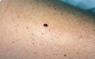
A 35-year-old woman was seen two months postpartum with a mole, which had enlarged during her pregnancy, on her right thigh. The mole was longstanding and measured 6 mm in diameter (Figure 1). Dermoscopy showed a pigmented nodule with an asymmetrical outline including a flat peripheral component. There were numerous irregularly sized and shaped dark brown globules that coalesced and extended focally to the border. A patchy grey–white veil was present. Scattered blue–black dots could also be seen at one border (Figure 2). The surgical specimen showed large irregular nests of atypical pigmented melanocytes both within the epidermis and underlying dermis, with a lymphocytic infiltrate (Figure 3).
Single article purchases are temporarily unavailable due to site maintenance.
If you would like to purchase an article during this time, please email us at [email protected] with the article details and we'll assist you directly. We'll also let you know when online purchasing is available again.
Thank you for your patience and understanding.