A new pigmented lesion
Melanoma
Skin cancer
Case presentation
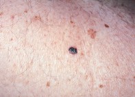
Over a six-month period, a 56-year-old man developed a new pigmented lesion, 0.8 cm in diameter, over his right lateral thigh (Figure 1). Dermoscopy revealed a dark asymmetrical lesion with multiple components and colours. The border was associated with irregular, blunt, pigmented projections (pseudopods). There were irregularly sized pigmented globules and dots that extended to the periphery and a prominent milky veil (Figure 2). Excision biopsy showed large nests of atypical melanocytes within the epidermis and upper dermis. Some of the nests were fused together and were associated with lymphocytic inflammation and pigment release into the surrounding dermis (Figure 3).
Single article purchases are temporarily unavailable due to site maintenance.
If you would like to purchase an article during this time, please email us at [email protected] with the article details and we'll assist you directly. We'll also let you know when online purchasing is available again.
Thank you for your patience and understanding.