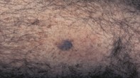
A mottled blue–grey lesion
Melanoma
Skin cancer
Case presentation
Over a six-month period, a 73-year-old man noticed that a longstanding tan-coloured lesion on the posterior aspect of his left calf had changed in colour (Figure 1). The lesion measured 1.5 cm in diameter. Under dermoscopy, it had a mottled blue–grey colour, with a patchy pale veil. There were pigmented dots and globules but no well-developed pigment network (Figure 2). Skin biopsy revealed an irregular epidermis with underlying superficial lymphocytic inflammation and melanin pigment. There was no melanocytic proliferation or nests present within the junctional zone (Figure 3).
Single article purchases are temporarily unavailable due to site maintenance.
If you would like to purchase an article during this time, please email us at [email protected] with the article details and we'll assist you directly. We'll also let you know when online purchasing is available again.
Thank you for your patience and understanding.