A crusty crimson plaque
Melanoma
Skin cancer
With sufficient training and expertise, clinicians can use dermoscopy to improve diagnostic accuracy for melanocytic lesions and other common skin tumours.
Case presentation
A 66-year-old woman presented with an asymptomatic red scaly patch on her leg that had been slowly enlarging over a two-year period. She was a renal transplant recipient and had a history of treatment for multiple nonmelanoma skin cancers affecting both legs.
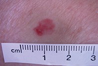
On examination, a well demarcated but irregularly-shaped scaly red plaque (9 mm diameter) was observed on the anterior aspect of the right shin (Figure 1). Areas of superficial ulceration were present.
Dermoscopy revealed subtle hyperkeratosis, multiple glomerular vessels and multifocal hypopigmentation (Figure 2). The absence of a discernible pigment network was significant.
Single article purchases are temporarily unavailable due to site maintenance.
If you would like to purchase an article during this time, please email us at [email protected] with the article details and we'll assist you directly. We'll also let you know when online purchasing is available again.
Thank you for your patience and understanding.