Prostate cancer detection. Advancing best practice
Progress in improving diagnoses for men with aggressive prostate cancer is occurring rapidly on many fronts, not least with regard to early detection of disease and disease recurrence.
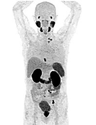
Australia is a world leader in the diagnosis of prostate cancer. Clinical guidelines used worldwide recommend a standard process for diagnosing prostate cancer that comprises prostate-specific antigen (PSA) blood testing followed by a random transrectal ultrasound-guided (TRUS) prostate biopsy procedure if the PSA test result is elevated.1 For many reasons this process is no longer best practice, and Australian practice is evolving ahead of the guidelines. Australian urologists and oncologists are adopting new imaging techniques, including prostate-specific membrane antigen positron emission tomography (PSMA PET) scanning (Figure 1), which is discussed below.
The problems with current standard practice
The first drawback of the current standard process relates to the limitations of PSA testing. An abnormally elevated PSA level can potentially have three causes: benign enlargement of the prostate, prostatitis or prostate cancer. About half of the men in the target age group for prostate cancer testing (men aged between 50 and 70 years) who have an elevated PSA level will not have prostate cancer.2 If all those with an elevated PSA level underwent a transrectal biopsy, 50% would be being subjected unnecessarily to an invasive procedure and the associated risk and harms.
A standard TRUS prostate biopsy is risky, inaccurate and, often, painful. In a TRUS biopsy, the biopsy needle is randomly passed through the wall of the rectum numerous times to take samples of the adjacent prostate gland. Faecal bacteria are commonly injected by the biopsy needle directly into the prostate gland, potentially causing acute bacterial prostatitis and, because of the rich blood supply to the prostate, septicaemia. Although it is an exceedingly rare outcome, men have died from septicaemia after TRUS biopsy of the prostate.3
Aiming to reduce this risk, urologists have been routinely prescribing broad-spectrum antibiotics for patients who undergo a TRUS biopsy. However, the typical preventive antibiotics used are becoming less and less effective as antibiotic resistance rapidly increases.4 Rates of septicaemia after TRUS biopsy are increasing around the world, and what should be a simple diagnostic procedure is actually dangerous.
When a TRUS biopsy is performed, the urologist has no idea where the cancer is or if cancer is present at all. Although ultrasound is used to guide the biopsy needle, this is simply to direct the needle into the prostate and it does not show where cancer might be located within it. It is therefore essentially a blind biopsy, which is why this technique misses clinically significant prostate cancer up to 30% of the time.5
Further, depending on what type of anaesthetic is provided, a transrectal biopsy is often painful and many men find it is embarrassing and anxiety provoking.6
Based on an initial 50/50 chance that a patient does not have prostate cancer; and if he does have prostate cancer there is a 30% chance of not detecting it; plus a 2% chance he will develop septicaemia;3 and the recommended standard investigation process is invasive, dangerous, inaccurate, and possibly painful and emotionally stressful … would you advise him to undergo this process?
Changing practice in Australia
This is where the bad news ends and the good news begins. In Australia, this so-called standard practice is gradually being consigned to history.
Multiparametric MRI
MRI (Figure 2) is a true game-changer in the diagnosis of prostate cancer, and Australian urologists have been early adopters of it.7 Using a standardised method of MRI, multiparametric MRI (mpMRI), clinically significant prostate cancer can be reliably detected. Strong high-level evidence now indicates that mpMRI should be recommended as the next step after finding a patient has an elevated PSA level.8-10 Prostate MRI should only be ordered by the treating urologist or other oncology specialist.
In contrast to a TRUS biopsy as the initial investigation after a raised PSA result, a prostate mpMRI is safe and painless. The only drawbacks associated with it are that some patients feel claustrophobic inside the MRI machine, and there is not yet a Medicare rebate, so the full cost of $400 to $1000 must be borne by the patient.
mpMRI scanning in patients who have an elevated PSA level can help the urologist decide whether a biopsy is needed at all. If the mpMRI is negative for prostate cancer, it is highly likely that there is no clinically significant prostate cancer present, and a biopsy can often be avoided. If the mpMRI does show a suspicious lesion, the subsequent biopsy can target it with high diagnostic accuracy. In summary, using MRI this way results in fewer unnecessary and invasive biopsies, but when a biopsy is needed, it is far more accurate.
The effectiveness of mpMRI of the prostate, however, depends on meeting world standards for scanning as well as reporting by a radiologist who has a high level of specific experience in prostate MRI scanning.11 Clinical experience tells us that such experience is relatively rare at present. For this reason, my colleagues and I have created an online platform to train radiologists and urologists around the world to read prostate MRI accurately and will launch this program in the next few weeks.
Transperineal biopsy
In Australia, the recent rate of uptake of transperineal biopsy by urologists as the preferred method for prostate biopsy is unprecedented.7 This method has been shown to have a rate of infection of zero or near zero, which therefore removes the need to prescribe broad-spectrum antibiotics.12-14
Prostate-specific membrane antigen positron emission tomography
Urologists and oncologists in Australia are also adopting the use of PSMA PET scanning for detecting early metastatic prostate cancer. Until now, bone scintigraphy and CT scans have been the options available for locating secondary metastases. Both types of scan have low sensitivity and specificity for this purpose, and therefore have limited usefulness for guiding appropriate treatment for prostate cancer. PSMA PET scanning uses a gallium-68-labelled ligand that binds to the prostate-specific membrane antigen that is overexpressed on prostate cancer cells.
A growing body of evidence shows PSMA PET scanning is far superior to traditional imaging for determining the extent of prostate cancer in a patient once cancer has been diagnosed (Figure 1).15 However, because this type of scan is so new, outcome data are not yet available to confirm whether these scans lead to improved patient outcomes.
Conclusion
More accurate diagnostic information must point us in the direction of optimal management and risk minimisation for men with prostate cancer. It is crucial, however, that clinical trials and registries are undertaken so that higher level evidence can potentially confirm what is being observed in clinical practice. Only then will official guidelines be able to catch up to cutting edge practice. MT

