Keratinised nodules
Skin cancer
Skin lesions
Case presentations
Case 1
A 78-year-old woman presented with an enlarging pink nodule on her left shin that had appeared 12 months previously. It had started as a pink plaque and had progressively thickened. The patient had a past history of chronic sun exposure and multiple squamous cell carcinomas (SCCs).
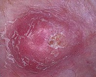
Examination revealed a firm and pale pink nodule measuring 9x10mm on the pretibial leg (Figure 1a). There was focal hyperkeratosis and the surrounding skin displayed evidence of solar damage. Polarised dermoscopy, performed using alcohol gel for immersion, revealed focal areas of keratinisation and diffusely distributed glomerular vessels (Figure 1b). Perivascular white halos were visible at the periphery. There were no pigmented structures.
The nodule was excised and histopathology confirmed a well differentiated invasive SCC.