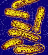
The changing face of investigating gut pathogens. Molecular testing
Digestive diseases
Diarrhoea due to enteric pathogens is a significant health burden. Molecular testing, especially multiplex polymerase chain reaction and microarray testing, is becoming routine for investigation of gut pathogens.
Diarrhoea due to enteric pathogens is a significant health burden. Data from the Bettering the Evaluation and Care of Health (BEACH) study of visits to GPs in 2015–16 revealed that diarrhoea accounts for 0.7% of encounters with GPs.1 Enteric infections are estimated to result in 2.71 million visits to doctors and half a million stool tests performed in laboratories each year.2 Enteric infections are often self-limiting but some patients require diagnostic investigations.
Traditional microbiological techniques that are used to detect intestinal pathogens, such as microscopy and culture, have limitations. Culture for common bacterial pathogens takes 24 to 48 hours, and antibiotic susceptibility testing takes up to 48 hours longer, so the practitioner must rely on syndromic treatment while awaiting results. Microscopy for parasites detects a broad spectrum of pathogens, but requires great expertise and lacks sensitivity. Commonly used methods for faecal virus detection are reasonably rapid and accurate but lack sensitivity, have low sample throughput and cover a narrow spectrum of gastrointestinal viruses.3
Molecular detection of pathogen nucleic acid (DNA or RNA) has increasingly become routine for microbiological investigation of gut pathogens, with multiplex polymerase chain reaction (PCR) and microarray testing forming the mainstay of methods. This has improved time to obtaining a result, and automation means the laboratory is better able to handle a high throughput of specimens. These tests can be performed by staff without highly specialised skills and thus are suitable for 24-hour availability. Increased access to more rapid results has sometimes led to an increase in requests for testing, but this may not be warranted, due to the largely self-limiting nature of intestinal infection.3 Some understanding of the benefits and limitations of the technology can improve the ability of doctors to request and interpret results of diagnostic tests.
Treatment algorithms for bacterial diarrhoea in Australia recommend empirical therapy and microbiological testing for patients with severe disease.4 Testing is not recommended for patients with milder disease, unless the patient is immunocompromised. Evidence is lacking for the effect of early or expanded faecal pathogen test results on patient outcomes. However, in the era of increasing antimicrobial resistance, most relevant to Shigella and Campylobacter species in the Australian setting, improved sensitivity and faster results allow tailored empirical therapy. Public health notification of Shigella results allows a more timely response to this highly infectious pathogen.
Molecular tests are, in general, more sensitive than culture or microscopy-based testing, with one study showing an increase from 53 to 75% of cases with pathogens detected.3 However, tests for certain pathogens have a tendency to result in higher rates of false positives, may detect carriage rather than disease or may detect organisms that are usually commensals. This can lead to situations in which a laboratory detects an organism for which a test was not requested by the doctor (e.g. when bacterial gastroenteritis was suspected but a commensal parasite is detected) and can lead to unnecessary antibiotic treatment and psychological distress.5 In addition, certain pathogens, notably Salmonella species and Entamoeba histolytica, may have a lower detection rate with molecular assays compared with traditional culture or microscopy.2,3
It is also important to note that multiplex PCR and microarrays are aimed at the more common pathogens detected in populations. If an unusual pathogen is suspected or the patient has travelled overseas, it may be worth discussing the requested tests with the microbiologist, because specific phenotypic or serological methods may be required. It is important to note that a negative result refers only to the pathogens included in the assay. Phenotypic testing with microscopy and other techniques may be required for detection of uncommon pathogens, especially uncommon parasitic infections.
Molecular testing also has implications for public health surveillance of outbreaks and rates of disease. Increased sensitivity of testing may mean cases are easier to detect and disease incidence is higher compared with earlier periods. Culture of faecal specimens with bacterial pathogens detected by molecular methods (‘reflex culture’) is still required to inform public health and foodborne disease surveillance programs.3
Quality assurance programs
A quality assurance program is an essential part of delivering accurate results to clinicians, and all accredited laboratories in Australia are expected to participate in them. Newer tests, such as molecular tests for enteric pathogens, have been adopted by diagnostic laboratories before quality assurance programs for these have become readily available. The Royal College of Pathologists of Australasia (RCPA) is pilot-testing a quality assurance program for molecular detection of enteric bacterial pathogens, and a pilot quality assurance test for molecular parasite testing began at the end of November 2017.
Sample preparation and nucleic acid extraction
Faeces is a complex sample matrix, with large numbers of bacterial, viral and fungal organisms, human cells, foreign organic material and bile salts. These substances can inhibit the PCR test, leading to an indeterminate result.
Nucleic acid extraction is complex and differs for different pathogens. Assays for viruses require viral RNA and DNA, and RNA is more fragile than DNA. Parasites have thicker cell walls and require physical as well as chemical methods of nucleic acid extraction.
Poor nucleic acid extraction efficiency, or overzealous methods that damage DNA, can lead to false-negative results. An internal control is recommended, which is often viral DNA from another species (e.g. equine herpesvirus) added (‘spiked’) into each sample to ensure that DNA present in the sample is detected in the assay, and not destroyed in the process of extraction.3
Multiplex PCR
Multiplex PCR refers to a nucleic acid detection assay in which multiple targets are included in one sample tube. Tests may be developed in the laboratory or commercially available, and may include targets for the more common pathogens, with a separate assay for rarer pathogens, or both. Separate multiplex PCR tests are available for bacteria, parasites or viruses. A higher number of targets leads to greater difficulty avoiding technical problems in assay design, with three to five targets usual for most assays. Commercially available assays use separate tubes or nested PCR techniques to cover a broad range of pathogens.2
Microarrays
Microarrays are chip-based assays with DNA targets attached; a large number of targets for multiple pathogens can be included. A single chip has thousands of oligonucleotide probes attached. These short DNA probes hybridise with pathogen nucleic acid extracted from the stool sample and labelled with fluorescent markers. The pattern of fluorescence is interpreted to detect pathogens.6 Commercial assays are available that detect viral, bacterial and parasitic causes of diarrhoea in a single test.
In a study of paediatric diarrhoea, standard-of-care tests ordered by doctors had a 29.7% positivity rate and the commercial microarray assay detected a potential pathogen in 69.9% of cases.7 Two analytes were detected in 25.2% of cases and 9.5% of cases had three or more analytes detected. In the absence of an accepted gold standard, the doctor must interpret multiple positive results in the clinical context. They may represent true false positives (which may be caused by nonspecific binding to targets); biological false positives (in which the organism is present but not causing disease), which constitute the vast majority of these positive results, particularly for Blastocystis or Dientamoeba; or genuine copathogens.
Diagnosis of acute gastroenteritis
Clinical distinction between bacterial and viral gastroenteritis is difficult. Both are usually self-limiting illnesses. Diagnosis of viral gastroenteritis can be important in institutional outbreaks, such as in daycare centres for young children, hospitals or aged care homes, or workers in such facilities. Cruise ships have also been the setting for norovirus outbreaks.8 Diagnosis can also help assess vaccine program efficacy, in the case of rotavirus (Figure 1).9 Laboratories do not always automatically test for viral infections in people older than 5 years, so if the diagnosis is important for reasons of infection control, this should be noted on the request form.
Molecular assays for viral enteric pathogens are more sensitive than traditional enzyme immunoassay (EIA) rapid tests, and allow for higher sample throughput, which is useful in outbreaks. Molecular assays also include multiple viral targets, whereas EIA tests usually include tests for one or two viruses. Children can excrete pathogenic viruses such as rotavirus and norovirus at low levels for prolonged periods, which calls into question the utility of molecular detection in children. However, asymptomatic people who are shedding virus usually have a lower viral load than infected people. Laboratories are able to determine carriage states with lower viral loads from the ‘crossing threshold’ (the number of PCR amplification cycles it takes for the pathogen to be detected). This may be used to determine a cut-off to distinguish true positive results from carriage.3
Bacterial gastroenteritis is often self-limiting, but treatment is usually required for shigellosis and for severe illness caused by other bacterial pathogens. Multiplex PCR and microarray assays are currently in use by several Australian laboratories. The assays differ in the spectrum of pathogens covered. Pathogens may include Salmonella, Campylobacter and Shigella/enteroinvasive Escherichia coli (EIEC)/Shiga toxin-producing E. coli (STEC). Other assays in routine use in Australian laboratories that participated in the RCPA quality assurance pilot program in 2017 include these pathogens plus various combinations of Clostridium difficile and less common pathogens such as Aeromonas, Plesiomonas, Yersinia enterocolitica, Listeria, E. coli O157, enteroaggregative, enteropathogenic and enterotoxigenic E. coli, and Vibrio species.10 Clinicians need to be aware that if a patient’s stool test is negative for bacterial pathogens, the spectrum of pathogens in the assay should be assessed and additional testing requested if clinically necessary. Any relevant information such as recent travel, seafood consumption, aquatic contact, recent antibiotic use or pregnancy, should be provided on the request form to assist the receiving laboratory in determining appropriate testing.
Detection of STEC in a timely manner may improve patient outcomes because early diagnosis of haemolytic uraemic syndrome and initiation of intravenous fluid resuscitation are beneficial.11 STEC may be missed by routine laboratory methods, and molecular screening will pick up milder cases that may be missed by traditional protocols. Atypical STEC strains have been responsible for large outbreaks of acute gastroenteritis including the South Australian mettwurst-associated outbreak.3
It is important that laboratories continue to perform reflex culture for samples with a positive PCR result for a faecal pathogen, as currently recommended by the Centers for Disease Control and Prevention.12 Some patients will require antibiotic treatment, and antibiotic resistance is becoming more common in Salmonella, Shigella and Campylobacter infections in Australia. In addition, foodborne disease surveillance still relies on culture for detecting outbreaks, and all Salmonella, Shigella and other foodborne disease isolates are sent to reference laboratories for typing. This allows the relatedness of isolates to be determined in order to pinpoint the source of an outbreak.
Campylobacter colitis
Campylobacter species cause a colitis that is often self-limiting. It is usually acquired from contaminated food, often poultry. Severe or persistent cases can require antibiotic treatment. Complicated infection can occur in the immunocompromised patient, such as bacteraemia or prolonged diarrhoea, and treatment may be required. Extraintestinal manifestations such as reactive arthritis and Guillain–Barré syndrome are also associated with Campylobacter infection. There are multiple Campylobacter species, and C. jejuni and C. coli are the most common in humans.
Culture for Campylobacter requires 48 hours of incubation under specialised conditions; thus molecular methods provide a more timely result.3 PCR testing for Campylobacter results in a higher rate of positive tests than culture; some of these may be false positives, but some probably reflect a rate of culture-negative, PCR-positive true disease.13,14 Reflex culture can help confirm the diagnosis. Ciprofloxacin resistance is becoming more common in Campylobacter isolates, especially those isolated from patients who acquired the disease overseas.
Salmonella gastroenteritis
Salmonellosis is often self-limiting, but may cause severe or prolonged infection. Standard culture methods for Salmonella have similar sensitivity to molecular detection.3,14 There is an increasing rate of antibiotic resistance in Australian nontyphoidal Salmonella isolates, although it is still very low, with 0.6 to 1.9% found to be ceftriaxone-resistant and 0 to 1.1% found to be ciprofloxacin-resistant.15 Reflex culture is required for susceptibility testing and serotyping for outbreak detection.
Shigella infection
Shigellosis can cause severe dysentery, as well as mild-to-moderate colitis. Shigella is highly infectious, and treatment is recommended both to treat disease and to reduce infectivity. Shigellosis may be sexually transmitted, especially when there is faecal–oral contact.
Antibiotic resistance is increasing in Shigella species. In a recent study, 35% of Shigella isolates in New South Wales were resistant to ciprofloxacin,16 and these authors have recommended azithromycin as a first-line treatment for suspected shigellosis. However, it should be noted that there have been outbreaks of multidrug-resistant Shigella in the men who have sex with men (MSM) community in Australia recently, which have been linked to overseas travellers; the resistance was to azithromycin.17
Molecular assays for shigellosis may detect the ipaH gene, which is also found in enteroinvasive E. coli (EIEC), and these do not distinguish between the two organisms. This can be done by means of reflex culture, but the illness is clinically indistinct. Molecular testing for Shigella detects substantially higher rates of disease than culture.14
Clostridium difficile infection
Molecular detection has excellent sensitivity for C. difficile infection (CDI) and is recommended as a first-line screening test for laboratories. This is best followed by a confirmatory assay for toxin production such as EIA, because some patients who test positive for C. difficile on PCR testing do not have clinical disease, and the positive PCR test may reflect asymptomatic colonisation with a toxigenic strain. EIA has imperfect sensitivity, however, so a negative EIA with a positive PCR test needs to be interpreted in the clinical context, i.e. some of these patients have CDI and some are asymptomatic carriers. CDI is extremely rare in children under 2 years of age, and laboratories should not issue results on young children, as the carriage rate is high.18
Culture of specimens with positive results is recommended for surveillance purposes.
Gastrointestinal protozoa
The most common intestinal parasitic infections in Australia are diarrhoeal illnesses due to the protozoan parasites Giardia, Cryptosporidium and Entamoeba histolytica (Figure 2). Infections with other intestinal parasites are relatively rare. Therefore, most molecular assays used in Australian settings include these three pathogens. Many assays also include Dientamoeba fragilis and sometimes Blastocystis species, which are the most common parasites found in stool samples in Australian populations.2 In the vast majority of children and adults, these two organisms are commensals. The RCPA has issued a statement which recommends that laboratories consider a multiplex platform that does not include these targets, and testing for them should be conducted only as requested by the clinician.
Traditional parasitology requires highly trained scientists for it to be performed well. Microscopy is reasonably rapid, but is labour intensive and throughput is low. It also lacks sensitivity when compared with molecular assays. Multiple stool specimens can increase sensitivity, but patient compliance with these requests is low. An additional logistical barrier is that D. fragilis in particular requires fixation of stool specimens at the time of collection, because the organism degenerates rapidly.2 Microscopy is still required to detect the broader range of parasites of clinical concern in populations such as Indigenous Australians and Australians who live remotely, travellers, refugees and patients who are immunocompromised.
Depending on the organism, molecular detection of parasites tends to be more sensitive than traditional methods. A comparative study conducted in a large pathology service in Queensland showed an increase from 11.4 to 31.5% of stool samples with parasites detected using molecular methods. Giardia was detected in 3.8% of samples when molecular detection was used, compared with 2.2% when traditional methods were used, and detection rates for Cryptosporidium were somewhat increased. A small number of E. histolytica infections were detected via traditional methods (four patients) and none were detected by PCR testing. The largest increase was for Dientamoeba, from 1% detected by microscopy to 17.6% detected by PCR testing. Blastocystis was also detected in 17.5% of specimens using PCR testing, compared with 7% by microscopy. Of the positive specimens, 24% had more than one parasite detected, with 74% of these being Blastocystis and Dientamoeba together.
This large increase in detection rates, and the high rate of co-detection with molecular methods, raise the possibility of many more patients being diagnosed with parasitic infection and treated with antibiotics. However, the true prevalence of disease caused by Blastocystis and Dientamoeba is much lower. It can be difficult to distinguish between disease and commensal carriage of protozoan parasites, especially Blastocystis and, to a lesser extent, Dientamoeba.
The significance of finding Dientamoeba in stool specimens has been debated since this organism was first described almost 100 years ago. Parasite eradication has been associated with symptom clearance in several prospective studies.2,19,20 Dientamoeba, uniquely among the protozoan intestinal parasites, may be associated with peripheral eosinophilia that resolves with treatment and parasite eradication.19,21 However, asymptomatic carriage and parasite persistence after therapy have also been documented.2,19 Byrne and Robson, in a large study in a community setting in Queensland, found that 54% of children aged 5 to 10 years had Dientamoeba in their stools.2 For most patients, treatment is not required.
Similarly, 17.5% of children in the same study had Blastocystis detected by molecular methods, which represented a large increase when compared with detection by microscopy.2 Blastocystis is a commensal organism with a worldwide distribution. It has been suggested that certain subtypes may be more likely to cause disease, but this remains unproven, and molecular assays currently in use do not distinguish between subtypes.22,23 Blastocystis found in a stool specimen should be considered a commensal organism.
Strongyloidiasis
Strongyloides stercoralis, a soil-transmitted nematode, can cause prolonged latent infection. It is unique among gastrointestinal parasites because its life cycle is completed within the human host, allowing infection to persist for decades after acquisition. It is endemic to the warmer northern parts of Australia and to other warm climates.
This helminth is of increasing importance in the era of increased immunosuppression. Reactivation of latent infection, particularly when a patient is being treated with corticosteroids, can lead to hyperinfection syndrome, which has an extremely high mortality rate.24
All diagnostic methods for strongyloides have limitations.25 Detection of larvae by microscopy is hampered by intermittent shedding and the usually low intensity of chronic infection. There are no ova or cyst forms to be found with microscopy. Stool culture is laborious, requires specialised methods and is a safety hazard. Serological testing is the best method for detecting latent infection, but results can be negative in elderly or immunocompromised patients. Molecular detection in stool is a useful adjunct to traditional methods, and is probably more sensitive in active infections, but its performance in latent infection is not yet well described. Its sensitivity is poorer than serological tests for latent infection, according to data available to date.25 Laboratory-developed molecular assays are available in some referral laboratories in Australia.
Molecular tests will continue to improve and become the routine method used by laboratories, so it is important to understand their limitations
Other intestinal parasites and microsporidia
Numerous other intestinal parasites may cause disease that can present with gastrointestinal symptoms, anaemia or hepatobiliary problems. These parasites include the roundworm Ascaris lumbricoides, hookworms Ancylostoma duodenale and Necator americanus, liver flukes Fasciola and Clonorchis sinensis, tapeworms Taenia solium and T. saginatum, and blood fluke Schistosoma mansoni, rarer protozoa such as Cyclospora and Isospora belli, and intestinal microsporidia. None of these is common clinically in Australia.
Molecular detection of these pathogens is not generally available. Testing for Cyclospora is included in some commercial multiplex PCR assays and the commercial microarray assay. It is important to note that these organisms will not be detected by laboratories using standard assays for their routine stool parasitology work. If the patient has peripheral eosinophilia, significant immunocompromise, a history of travel or other atypical features, the case should be discussed with the clinical microbiologist to ensure appropriate traditional parasitology methods are employed.
Conclusion
Molecular assays for enteropathogens offer some important advantages to the clinician in terms of rapid results and improved sensitivity for most targets. However, there are some clinical challenges. The possibility of a negative result when the causative pathogen is not included in the panel needs to be considered at all times. Improved sensitivity corresponds to an increased rate of false-positive results. Improved sensitivity also means that carriage states, of both commensal organisms and pathogens, are more likely to be detected. Therefore, all results should be considered in the clinical context.
Reflex culture for positive results is still needed for antibiotic treatment and public health purposes. Molecular tests will continue to improve and become the routine method used by laboratories, so it is important to understand their limitations. MT