Out cold: cryotherapy for skin lesions
Skin cancer
Skin lesions
Cryotherapy is an important tool used by many GPs in treating skin lesions. A diagnosis is essential before freezing a lesion, and cryotherapy is most effective for superficial lesions.
Many general practitioners and most dermatologists use cryotherapy every day to produce quick, effective results with usually only minor discomfort to the patient. The use of cold to treat skin lesions dates from 1899, when Dr A.C. White, a New York dermatologist, dipped a cotton-tipped applicator in liquid air and successfully treated warts, keratoses and other lesions.1 Liquid nitrogen spray was introduced in the 1960s by Dr Setrag Zacarian, a dermatologist in Boston.1 Liquid nitrogen has a boiling point of -196°C and is still the preferred cryogen.
Uses of cryotherapy
Many texts and articles on cryotherapy (or cryosurgery) have advocated its use for nearly every imaginable skin lesion, from inflammatory dermatoses to malignant tumours.1-3 The best results are when freezing is used on types of lesions for which its efficacy has been proven.
In general terms, cryotherapy is most effective for superficial lesions. For deeper lesions, the response may be only partial, morbidity after treatment may be considerable, healing can be slow, and the resultant hypopigmented depressed scar may be unacceptable. Types of lesions commonly treated with cryotherapy are listed in Box 1.
Supply and storage of liquid nitrogen
A vacuum flask can be filled with liquid nitrogen at a supply depot, or more commonly from a Dewar flask kept at the doctor’s office. The Dewar flask is filled as needed by a liquid nitrogen supplier for a moderate cost (less than $2.00 per litre plus a delivery charge and rental charge for the flask unless the practice owns one), and its large volume helps to ensure a ready supply is always on hand. Efficient vessels have holding times of up to 90 days.
A vacuum flask containing liquid nitrogen must never be capped unless the cap is perforated to allow escape of gaseous nitrogen. Capped flasks may otherwise explode.
Methods of application
Two methods are commonly used to apply liquid nitrogen to skin lesions:
- a cotton-tipped applicator
- a handheld spray device (‘gun’).
Cotton-tipped applicators rolled by hand are better than proprietary ones, as the latter are tightly rolled and do not hold nitrogen well. A portion of cotton wool is teased out and rolled onto an orange stick with a twirling motion between the index finger and thumb (Figures 1a and b). This produces a large applicator suitable for most lesions. Applying the nitrogen in this way is particularly useful for lesions on the face, especially around the eyes, as many patients do not like nitrogen sprayed in that area.
When a cotton-tipped applicator is used, the liquid nitrogen should be decanted into a styrofoam cup or small steel bowl, to avoid contamination of the nitrogen container. This applies especially when treating viral warts, but it is a prudent measure for all lesions.
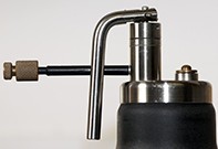
Several models of nitrogen gun are available that vary in capacity and other features. The unit may need to be refilled two or three times a day from the Dewar flask if it is being used regularly. Spray tips with different apertures to regulate the flow of nitrogen are supplied with the device, as well as various probes and nozzles for difficult sites or unusual applications.
The procedure
Cryotherapy involves tissue destruction. The mechanism is divided into four phases as follow.4
- Heat transfer. Heat is transferred rapidly from the tissue to the cryogen.
- Tissue injury. Ice crystals form, initially outside cells but then intracellularly, damaging cell membranes and intracellular organelles permanently.
- Vascular stasis and occlusion. Dermal vasculature is damaged, leading to ischaemic necrosis of treated tissue.
- Inflammation. Over the next day, oedema and inflammation follow, and healing occurs over successive days.
Healing times vary with the site, degree of freezing and the patient’s general health. More damage is done with longer freezing times and repeated freeze–thaw cycles. The time taken to thaw is an indication of the degree of tissue damage.
If a cotton applicator is used, it is dipped into the nitrogen and then applied firmly to the lesion. The time of application will vary according to the lesion type; your experience will determine the appropriate application time. If a nitrogen dispenser with a large nozzle aperture is used, then a spray time as short as two seconds may be all that is required. If a finer spray nozzle is used, the same lesion may require a longer spray.
Some cells are more sensitive to cold; melanocytes are more sensitive than keratinocytes, explaining the varying frequencies of postinflammatory hypopigmentation at different cryotherapy sites. Thicker lesions require deeper and longer freezing, but this will increase side effects both in the short and long term.
A diagnosis is best made before cryotherapy is undertaken.5 If the diagnosis is not apparent then it should be obtained by referral or skin biopsy. The ‘let’s freeze it and see what happens’ approach has no place in today’s increasingly litigious society.
It is advisable for less experienced cryotherapists to initially treat only some of the lesions listed in Box 1 (principally actinic keratoses, small seborrhoeic keratoses and viral warts) and to otherwise refer the patient to a dermatologist.
Cryotherapy for different types of lesions
Actinic keratoses
In Australia, actinic keratoses are common. Lesions vary from pink, slightly scaly patches in exposed sites (Figure 2) to indurated horny nodules in more advanced lesions (Figure 3).
Actinic keratoses are precursors of squamous cell carcinoma. Signs of conversion to squamous cell carcinoma include pain, rapid growth and fissuring. Lesions where there is some suspicion of conversion should be biopsied or excised. Sites prone to metastasis from squamous cell carcinoma include the ear, lip and scalp.
Actinic keratoses are frequently infected, especially those on the scalp and lip. Staphylococcus aureus is the usual cause. Precryotherapy treatment with saline bathings or with a suitable antiseptic can settle the secondary infection and clarify the diagnosis, especially when early squamous cell carcinoma is suspected.6
The technique for freezing an actinic keratosis is to apply liquid nitrogen to the lesion (Figure 4) and then allow it to thaw. The time taken to thaw depends on the amount of freezing induced.
The lesion can be grasped between the index finger and thumb and moved over deeper tissues to assess the depth of freeze. The frozen lesion has the consistency of a credit card. Thinner actinic keratoses will respond to a two to three-second freeze time with the largest aperture nozzle on the gun and a 20-second thaw, but thicker lesions will need a five to 15-second freeze, which will result in a 40 to 60-second thaw. A second freeze and thaw may be required if the lesion is thick, as sometimes thicker lesions do not respond to one freeze. Edge recurrence is quite common in diffuse actinic keratoses (Figure 5). If a lesion persists after adequate cryotherapy, the diagnosis may need to be reviewed.
Viral warts
Liquid nitrogen cryotherapy is an effective treatment for many types of viral wart. Common warts may need several treatments spaced a few weeks apart (Figure 6 and Figure 7). Plantar warts may need many treatments, as well as adjunctive therapy with sclerosant pastes. Treatment of hand warts in children with liquid nitrogen is painful, and subungual warts usually do not respond. Alternative measures (such as wart paints under occlusive tape) may be preferable as first-line therapy for warts in children. Most astute toddlers will allow a practitioner only one attempt with liquid nitrogen therapy, so the modality is best reserved for small warts only.
When a cotton-tipped applicator is used to treat warts, it is applied firmly to the wart. As liquid nitrogen does not kill the human papillomavirus, the treatment of warts warrants the use of a new applicator for each patient and the decanting of liquid nitrogen into a styrofoam cup to avoid contamination of the main container. The decanted nitrogen should be discarded after treatment of each patient.
Seborrhoeic keratoses
Small seborrhoeic keratoses respond well to liquid nitrogen, and many lesions can be treated at one sitting with only minor discomfort. Larger and thicker lesions require longer freeze times and heal more slowly, and scarring may result. Such lesions (Figure 8 and Figure 9) are better treated by point electrodesiccation under local anaesthesia.
During point electrodesiccation, the lesion is anaesthetised and lightly hyfrecated. The charred lesion is wiped off with a gauze swab, often coming away cleanly because the heat causes a cleft between the lesion and the underlying dermis. If the char is stubborn, a gentle curette will loosen it.
Basal cell carcinoma
Superficial basal cell carcinoma in low-risk sites responds well to appropriate cryotherapy. Successful treatment requires experience.3 Recurrence rates are high for inappropriately treated lesions (Figure 10 and Figure 11). Most dermatologists today would not use cryotherapy for basal cell carcinomas on the head and neck. The Australian Cancer Network recommends that cryotherapy is best reserved for low-risk tumours. This generally means well-defined, superficial, basal cell carcinomas of the trunk and limbs (Figures 12a and b).5
Skin tags (acrochordons) and pedunculated seborrhoeic keratoses
A useful method for treating skin tags is the forceps-grip method. A pair of large nontoothed McIndoe forceps, DeBakey plain forceps or similar are dipped in liquid nitrogen that has been decanted into a styrofoam cup. After 10 seconds or so, once the forceps are cold, the skin tag is grasped at its narrowest point (the neck) until it becomes frozen. The forceps need to be held with gauze to prevent frostbite in the operator’s fingers. This procedure is less painful than other methods of application and is less likely to produce post-treatment hypopigmentation.
Pigmented lesions
Some pigmented lesions are suitable for cryotherapy, the most common of which is the small seborrhoeic keratosis. However, cryotherapy has no place in the treatment of naevi.5
Side effects of cryotherapy
Patients should be told how a frozen lesion is expected to respond (e.g. Figure 13). A patient information handout (Box 2) can reassure the patient and prevent unnecessary telephone calls to the doctor’s office but invite them to follow up if they are worried.
Side effects vary with the depth of freeze, the type of lesion frozen, the site and the general health of the patient. Box 3 gives a comprehensive list of side effects.5 Some of these would be expected only with very deep cryotherapy.7
Asian patients or those with olive skin may experience hyperpigmentation, which settles after a few months. Figure 14 shows hyperpigmentation resulting from cryotherapy for a plane seborrhoeic keratosis.
One of the most common and annoying side effects is hypopigmentation (Figure 11 and Figure 15). This is common, even with light cryotherapy. Hypopigmentation is most obvious in areas of freckling or poikiloderma, which may be most apparent on the chest and lateral neck. Hypopigmentation may be limited by repeated lighter freezes and by feathering the edge of a frozen area (that is, freezing the periphery more lightly than the centre of the lesion). MT