What’s the diagnosis?
A child with a reticulated erythematous rash


Case presentation
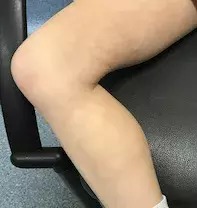
A 10-year-old girl presents with an asymptomatic, reticulated, erythematous rash on the medial aspects of her thighs and lower legs of one-week duration (Figure 1). Her parents report that the rash appears worse when she has been outside or running around. The girl is otherwise well and does not have a history of any skin diseases.
On questioning, the parents say that the girl had mild coryzal symptoms approximately two weeks earlier and they recall transient facial erythema. She was not febrile at any stage.
Differential diagnoses
Conditions to consider among the differential diagnoses for a child of this age include the following.
- Scarlet fever. This infection with group A streptococcus, most commonly seen in children, results in a generalised rash. It typically follows streptococcal pharyngitis or impetigo. A characteristic sandpaper-textured, erythematous rash develops over the neck, ears and trunk 12 to 48 hours after the acute onset of fever, headache, nausea, vomiting and anorexia. The patient is initially unwell but fever starts to resolve one to two days after the commencement of appropriate antibiotics. The timing of the rash and the mild preceding illness in the case patient described above indicate that this is not the correct diagnosis.
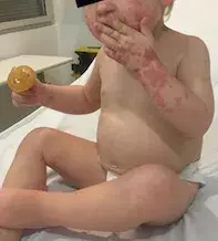
- Enterovirus infection. Non-polio enteroviruses are responsible for a wide range of viral exanthemata. Infection is most common in children and is spread by faecal–oral or respiratory routes. It is more prevalent in summer and autumn. The most common and most recognisable enterovirus exanthem is hand, foot and mouth disease; however, exanthemata can assume many and varying morphologies (Figure 2). Enteroviruses are important in the differential diagnosis of viral rashes and can also cause meningitis and encephalitis. The clinical picture in the case patient indicates a different aetiology.
- Rubella. The incidence of this RNA virus infection has decreased markedly since initiation of the measles–mumps–rubella vaccine. Similar to this case, an erythematous rash appears on the face spreading to the rest of the body about five days after a viral prodrome. However, the exanthem does not appear lacy or reticulated and the eruption typically fades after two to three days.
- Systemic-onset juvenile idiopathic arthritis. Also known as Still’s disease, this is an autoinflammatory condition typically associated with polyarthritis. It is estimated that 90% of children with acute febrile onset of Still’s disease have an exanthem.1 This eruption is erythematous, transient and coincides with the fevers. Although the rash is typically associated with arthralgias, it can precede joint pain by up to several years.
- Erythema infectiosum secondary to parvovirus B19 infection. This is the correct diagnosis, and is also known as fifth disease or ‘slapped cheek disease’. Parvovirus B19 is the only parvovirus known to affect humans. It was found to cause the typical features of erythema infectiosum in 1983.2 The virus most commonly affects children aged 4 to 10 years. Patients initially have mild prodromal flu-like symptoms (myalgia, headache and low-grade fever). The classic ‘slapped cheek’ appearance occurs seven to 10 days after the flu-like symptoms, with bright red erythema of the cheeks, sparing the nasal bridge. After this, a rash with a reticulated lacy pattern occurs on the extremities. It can remit and exacerbate for several weeks. The rash can worsen on exposure to sunlight or heat. Associated arthralgia, most commonly of the fingers, wrists and ankles, occurs more commonly in adults and can be prolonged.
Investigations
The diagnosis of erythema infectiosum secondary to parvovirus B19 infection is usually clinical, based on the progression of the rash from the ‘slapped cheek’ appearance to the reticulated exanthem. As well as the typical rash there are several established clinical associations with parvovirus B19, which may necessitate confirmation of the virus. These include polyarthropathy, papular-purpuric gloves and socks syndrome, transient aplastic crises in at-risk individuals (decreased red cell production) and hydrops fetalis. The risk of hydrops fetalis is greatest in the first 20 weeks of gestation. The overall risk of hydrops fetalis and stillbirth is low (3.9% and 0.6%, respectively) and pregnant women are not routinely screened for parvovirus B19 infection unless they have been exposed.3 If confirmation of infection is required – for example, in anaemic or immunocompromised children – serological testing for parvovirus IgG and IgM or parvovirus PCR will indicate the presence of infection. It should be remembered that the potential for virus transmission is early in the disease course, between the prodrome and onset of the rash.
Management
There is no specific treatment for erythema infectiosum secondary to parvovirus B19 infection. Affected children may remain at school as once the rash develops the virus is no longer contagious and they are typically not symptomatic. Red cell or intrauterine transfusion may be required for aplastic crisis or hydrops fetalis, respectively.
References
1. Calabro JJ, Marchesano JM. Rash associated with juvenile rheumatoid arthritis. J Pediatr 1968; 72: 611-619.
2. Heegaard ED, Brown KE. Human parvovirus B19. Clin Microbiol Rev 2002; 15: 485-505.
3. Enders M, Weidner A, Zoellner I, Searle K, Enders G. Fetal morbidity and mortality after acute human parvovirus B19 infection in pregnancy: prospective evaluation of 1018 cases. Prenat Diagn 2004; 24: 513-518.
Viral infections

