What’s the diagnosis?
A scaly brown rash on the lower legs


Case presentation
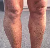
A 45-year-old man presents after noticing that the skin of his lower legs has become unusually scaly and brown during the winter months (Figure 1). The rash is not itchy.
The patient has a long history of ‘dry skin’, dating back to his early childhood. He mentions that his younger brother also has dry skin but his parents, sister and two daughters do not. Until a recent move to Victoria, the patient had lived in northern Queensland all his life.
Differential diagnosis
Conditions to include in the differential diagnosis include the following.
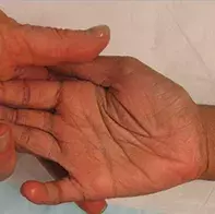
- Ichthyosis vulgaris. This is the most common and mildest form of the inherited ichthyoses, a heterogeneous group of genetically determined disorders characterised by dry, rough and scaly skin. Caused by a loss of function in the filaggrin gene, ichthyosis vulgaris is an autosomal-dominant condition with variable penetrance, affecting approximately 1 in 250 people, with males and females affected equally.1 Keratosis pilaris and hyperlinear palms and soles are present in most cases, and help to differentiate ichthyosis vulgaris from other forms of ichthyosis (Figure 2). Patients typically present with small, white and/or grey flaky scales that peel from the skin, and little to no itch. It is more commonly distributed on the extensor surfaces of the extremities, particularly on the legs, while the torso and flexural regions are usually not affected. There is a strong association with atopy: up to 50% of patients with ichthyosis vulgaris display features of atopic dermatitis, asthma or hay fever.2 Ichthyosis vulgaris is commonly mistaken for atopic eczema but is differentiated by a lack of itch and erythema. For the patient described above, ichthyosis vulgaris would be considered as a diagnosis, but typically the scales are not as brown in colour and more peeling is evident.
- Atopic dermatitis. Affecting up to 10% of adults, atopic dermatitis is a chronic, inflammatory skin condition characterised by pruritus and dryness. It commonly occurs in the flexural regions – such as inside the elbows and behind the knees – but it can affect any part of the body. Other clinical features include thickened skin, lichenification and excoriations. There are various causes and contributing factors: intrinsic factors include altered barrier function of the epidermis, defects in filaggrin function and immune dysregulation; extrinsic factors include irritants and allergens as well as an altered microbiome on the skin surface. The vast majority of cases appear in early childhood – many remit but a large proportion of patients continue to be affected into adulthood. It may also be associated with other atopic conditions, such as allergic rhinitis and asthma. It would be highly unusual for a patient with atopic dermatitis to have scaly skin without inflammation and no itch, as in this patient’s case. It would also be uncommon for it to worsen rather than improve with age.
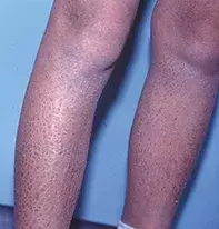
- X-linked recessive ichthyosis. This is the correct diagnosis. Occurring in 1 in 5000 males, X-linked ichthyosis is a gender-specific condition caused by deletion or mutation of the steroid sulfatase gene on the X chromosome. This leads to decreased production of cholesterol in the epidermis and an excess of keratinocytes, which is responsible for the characteristically scaly skin. The scales are typically large and brown in colour (Figure 3), and they predominantly appear on the abdomen, outer thighs and extensor surfaces of the upper arms and the legs. The scale is often described as being similar to fish scales (the term ichthyosis is derived from the Greek ‘ichthys’ for fish). There is little to no peeling of scales, and the palms and soles are not affected – these help to differentiate the condition from ichthyosis vulgaris. Newborn babies often present with a more generalised, fine scaling that becomes brown in colour during adolescence and adulthood. Patients with darker skin present with darker scales.
Diagnostic clues
When mild, X-linked recessive ichthyosis is commonly undiagnosed for a prolonged period or mistaken for ‘dry skin’, as in this case. Patients with ichthyosis who live in humid climates may be unaware of it. In this patient’s case, it was the change in his environment when he moved from Queensland to Victoria that led to the condition declaring itself for the first time. The fact that his younger brother has ‘dry skin’ and that his daughters and sisters do not are clues to a genetic X-linked condition. A proportion of patients with X-linked recessive ichthyosis have extracutaneous features, including testicular maldescent and asymptomatic corneal opacities.
Investigations
The most important features of the clinical history of a patient with X-linked recessive ichthyosis are the sex of the patient and a positive family history. Serum lipoprotein electrophoresis can show increased mobility of LDL-cholesterol, which is suggestive of X-linked recessive ichthyosis.3 The diagnosis can be confirmed through chromosomal analysis using fluorescent in situ hybridisation (FISH), which can detect the absence or mutation of the steroid sulfatase gene.
Management
X-linked recessive ichthyosis alone is usually a mild disease, with most patients managing their condition with regular application of emollients. Sun exposure, humid environments and short, lukewarm showers improve symptoms; avoidance of products that dry out the skin is also beneficial. The scaly appearance of the skin can be reduced with keratolytic agents such as urea and salicylic acid. A commercial mixture of topical urea, lactic acid and sodium pyrrolidone carboxylate is available that is often effective; however, it may be more suitable for use in older children and adults than babies and small children because it can cause stinging. In severe cases, there may be a role for topical retinoids to reduce the scaly appearance of the skin, but this may cause more skin dryness.
It is important to be aware of X-linked recessive ichthyosis because in female carriers it can result in prolonged labour associated with low maternal oestriol due to placental steroid sulfatase deficiency. Female carriers are phenotypically normal, but corneal stromal deposits may be detected on slit-lamp examination.
References
1. Smith FJ, Irvine AD, Terron-Kwiatkowski A, et al. Loss-of-function mutations in the gene encoding filaggrin cause ichthyosis vulgaris. Nat Genet 2006; 38: 337-342.
2. Osawa R, Akiyama M, Shimizu H. Filaggrin gene defects and the risk of developing allergic disorders. Allergol Int 2011; 60: 1-9.
3. Arndt T1, Pelzer M, Nenoff P, et al. Lipoprotein and apolipoprotein electrophoresis in X-chromosome recessive ichthyosis [article in German]. Hautarzt 2000; 51: 490-495.
Genetic disorders

