Peer Reviewed
Perspectives on dermoscopy
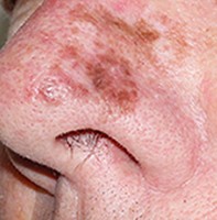
A pigmented macule on the nose: what is your diagnosis?
An updated version is available in the
linked supplement
Recent articles on:
Skin cancer
Skin cancer
Recent articles on:
Skin lesions
Skin lesions
Abstract
The differential diagnosis of pigmented macules of the face can be challenging. Dermoscopy may help, and adding confocal microscopy improves sensitivity and specificity; histopathology, however, remains the gold standard.
Key Points
Case presentation
A man in his 60s presented for a full skin check. He had heavily sun-damaged skin and a past history of lentigo maligna on the nose that was treated three years ago with cryotherapy and imiquimod. He had noted some new pigmentation arising in that area. On clinical examination, an irregular pigmented macule of two colours was seen (Figure 1). Differential diagnoses included solar lentigo, flat seborrhoeic keratosis, pigmented actinic keratosis and, most importantly, recurrent lentigo maligna.