Recurring pigmentation in an old scar
Skin conditions
Case presentation
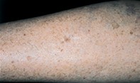
A 70-year-old woman had an 8 mm diameter mole removed from her left leg, and the mole was diagnosed histologically as a dysplastic junctional naevus. Eight years later, an irregular pigmented patch measuring 6 mm in diameter was noted at the lower border of the surgical scar (Figure 1). Dermoscopy revealed an asymmetrical, ill defined, broken pigment network with scattered small dark dots and isolated brown globules (Figure 2). Excision biopsy showed confluent proliferation of small hyperchromatic melanocytes in the junctional zone and focal collections of atypical melanocytes in the upper dermis (Figure 3).
Single article purchases are temporarily unavailable due to site maintenance.
If you would like to purchase an article during this time, please email us at [email protected] with the article details and we'll assist you directly. We'll also let you know when online purchasing is available again.
Thank you for your patience and understanding.