A young man with pruritic nodules on the genitals
Test your diagnostic skills in our regular dermatology quiz. What is the cause of these itchy nodules and widespread excoriations?
Case presentation
A 23-year-old man presents with an eight-week history of generalised pruritus affecting his whole body, with particular involvement of the genitals.
The itch is worse at night and has not responded to treatment with topical corticosteroids. He denies new sexual partners and is in a stable long-term relationship.
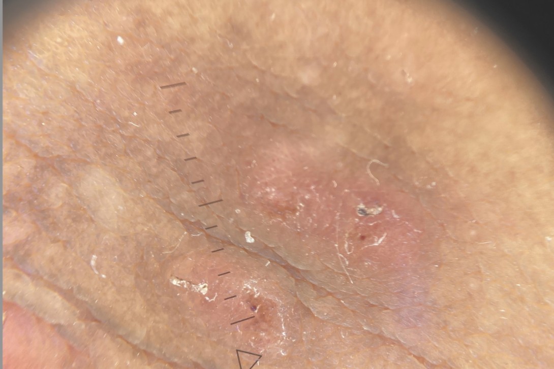
On examination, firm nodules are observed on the genitals, with widespread excoriations and minor involvement of the palms and fingers.
Differential diagnoses
Conditions to consider among the differential diagnoses include the following.
The case patient's lesions at presentation are shown in Figure 1.

Secondary syphilis
Syphilis is a sexually transmitted infection caused by the spirochaete Treponema pallidum.1 Secondary syphilis typically develops six to eight weeks after a primary chancre, which may have gone unnoticed. The characteristic presentation includes a widespread, symmetrical, nonpruritic maculopapular eruption that classically involves the palms and soles. This is often accompanied by condylomata lata (moist, flat, greyish-white plaques in intertriginous areas, including the genitals and perianal region), mucous patches, lymphadenopathy and constitutional symptoms such as fever, malaise and weight loss.1,2 The genital lesions seen in patients with secondary syphilis tend to be flat, moist plaques. The diagnosis is confirmed by serological testing, including treponemal and nontreponemal assays.2
Syphilis is an important consideration for a young man with genital lesions. For the case patient, however, the intensely pruritic nature of the eruption and the morphology of the lesions (firm nodules rather than flat condylomata) were not consistent with secondary syphilis.
Anogenital warts
Anogenital warts are caused by human papillomavirus (HPV), most commonly types 6 and 11.3 They present as soft, flesh-coloured, exophytic papules or pedunculated lesions on the genitals, perineum or perianal region. The lesions may be single or multiple and can coalesce into larger cauliflower-like masses. They are typically asymptomatic, although patients may report itch or discomfort.3,4 The diagnosis is usually clinical, based on the characteristic verrucous morphology. Biopsy is rarely required but may be considered if the diagnosis is uncertain or if there is concern about atypical features.4
For the case patient, a diagnosis of anogenital warts was considered. However, the genital lesions were firm and smooth, rather than verrucous or papillomatous, and the widespread pruritus with excoriations elsewhere on the body was not explained by HPV infection alone.
Hidradenitis suppurativa
Hidradenitis suppurativa is a chronic, relapsing, inflammatory condition of the apocrine gland-bearing skin, including the axillae, groin, gluteal and inframammary regions.5 It typically presents after puberty with painful, deep-seated nodules, abscesses, sinus tracts and scarring in the affected areas. The disease is characterised by a pattern of flares and remissions and may lead to significant morbidity and reduced quality of life.5,6 Risk factors include obesity, smoking and a family history of the condition. The diagnosis is clinical, based on the presence of typical lesions in characteristic locations with a history of recurrence.6
For the case patient, the genital nodules raised the possibility of hidradenitis suppurativa, but the nodules are typically tender and inflammatory, progressing to abscesses and sinus tracts, rather than firm, as observed in this case. Furthermore, the widespread pruritus and excoriations were not consistent with this diagnosis, which does not cause generalised itch.
Scabies
This is the correct diagnosis. Scabies is an infestation of the skin caused by the mite Sarcoptes scabiei var. hominis. It is a common condition, with over 200 million people affected worldwide at any given time.7 Transmission occurs primarily through prolonged, close, skin-to-skin contact, and sexual contact is an important route of transmission among young adults.8
The clinical hallmark of scabies is intense, generalised pruritus that is characteristically worse at night. The itch is caused by a delayed type IV hypersensitivity reaction to the mite, its eggs and faecal material (scybala), which typically develops two to six weeks after initial infestation.7,9 In reinfestation, symptoms may develop within one to three days because of pre-existing sensitisation.
The classic distribution of scabies includes the interdigital web spaces, wrists, antecubital fossae, axillae, periumbilical region, waistline, buttocks and genitals.8 In immunocompetent adults, the head and neck are usually spared. Examination findings include fine, linear or serpiginous burrows (the pathognomonic sign), small erythematous papules and excoriations from scratching.
Nodular scabies, a variant of the condition, is characterised by the development of firm, pruritic, red-brown nodules, typically 5 to 20 mm in diameter, that occur most commonly on the male genitalia (penile shaft, scrotum, glans), buttocks and axillary folds. These nodules represent a vigorous hypersensitivity response to retained mite antigens and may persist for weeks to months after successful eradication of the mite, even in the absence of active infestation.10
Diagnosis
The diagnosis of scabies is based on a combination of clinical features and, where possible, confirmatory investigations. The 2020 International Alliance for the Control of Scabies has developed consensus criteria for diagnosis of scabies according to level of certainty (Box).11 Skin scrapings taken from the end of a burrow and examined under light microscopy with mineral oil may reveal mites, eggs or scybala. Dermoscopy is useful and noninvasive – the characteristic finding is the delta-wing jet sign (also called the jet with contrail sign), which corresponds to the triangular, dark-brown anterior end of the mite at the leading edge of a burrow.12

For the case patient, a confirmed diagnosis was achieved through microscopic examination of skin scrapings, which revealed the presence of a scabies mite. Dermoscopy showed a short curvilinear whitish burrow with a subtle dark-brown, triangular or ovoid structure at the leading edge, consistent with the delta-wing jet sign of scabies (Figure 2).

Management
The management of scabies involves eradication of the mite and symptomatic treatment of pruritus. All household members and close physical contacts (including sexual partners) should be treated simultaneously, regardless of whether they are symptomatic, to prevent reinfection.13,14 Appropriate environmental measures should also be implemented.
Eradication therapy
Topical treatment
Permethrin 5% cream is first-line topical treatment for scabies in Australia.13 It is applied to the entire body from the neck down (including under the nails, between the fingers and toes, and on the genitals), left on for at least eight hours (usually overnight) and then washed off. A second application should be performed one week later to kill any newly hatched mites. All areas of skin should be treated, not just those with visible lesions.13,14 Patients should be advised to apply the cream to cool, dry skin; application immediately after a hot bath may increase absorption and reduce efficacy.
Benzyl benzoate 25% emulsion is an alternative topical agent; it is applied for 24 hours and then washed off, with a second application one week later. It is less well tolerated because of skin irritation.13
Oral treatment
Oral ivermectin (200 microg/kg, administered as a single dose, repeated after seven days) is an effective systemic treatment for scabies.15 It is particularly useful for patients who have extensive disease or are immunocompromised and those in whom topical treatment has failed. Ivermectin is also useful in outbreak settings where individual application of topical agents is impractical.13,15
Symptomatic management
Symptomatic relief of itch can be achieved with an oral antihistamine (e.g. cetirizine or loratadine) and regular application of emollients. A moderate-potency topical corticosteroid can be applied to persistent pruritic areas after confirmed eradication of the mite.13 Post-scabietic itch is common and may persist for two to four weeks after successful treatment, because of the ongoing hypersensitivity response.9 Patients should be counselled that persistence of itch does not necessarily indicate treatment failure.
Scabietic nodules may persist for several weeks to months after successful treatment.10 If nodules remain symptomatic, a potent topical corticosteroid (e.g. betamethasone dipropionate 0.05% ointment) applied under occlusion or intralesional triamcinolone acetonide (2.5 to 5 mg/mL) may be considered to hasten resolution.10
Environmental measures
Clothing, bed linen and towels used in the preceding three days should be washed in hot water and dried in a hot dryer or sealed in a plastic bag for at least 72 hours, as mites cannot survive off the human host for longer than this period.14
Outcome
The patient and his partner were treated with overnight topical permethrin 5% cream, which was repeated one week later. He experienced post-scabietic itch for several weeks, which settled with use of betamethasone dipropionate 0.05% ointment and emollients. He was also given advice about the importance of environmental measures to eradicate the mite at home. MT
COMPETING INTERESTS: None.
References
1. Hook EW 3rd. Syphilis. Lancet 2017; 389: 1550-1557. [Erratum in: Lancet 2019; 393: 986.
2. Kingston M, French P, Higgins S, et al. UK national guidelines on the management of syphilis 2015. Int J STD AIDS 2016; 27: 421-446.
3. Lacey CJ, Woodhall SC, Wikstrom A, Ross J. 2012 European guideline
for the management of anogenital warts. J Eur Acad Dermatol Venereol 2013; 27: e263-270.
4. Yanofsky VR, Patel RV, Goldenberg G. Genital warts: a comprehensive review. J Clinical Aesthet Dermatol 2012; 5: 25-36.
5. Jemec GB. Hidradenitis suppurativa. N Engl J Med 2012; 366: 158-164.
6. Ingram JR. Hidradenitis suppurativa: an update. Clin Med 2016; 16: 70-73.
7. Karimkhani C, Colombara DV, Drucker AM, et al. The global burden of scabies: a cross-sectional analysis from the Global Burden of Disease Study 2015. Lancet Infect Dis 2017; 17: 1247-1254.
8. Arora P, Rudnicka L, Sar Pomian M, et al. Scabies: a comprehensive review and current perspectives. Dermatologic Ther 2020; 33: e13746.
9. Walton SF, Currie BJ. Problems in diagnosing scabies, a global disease in human and animal populations. Clin Microbiol Rev 2007; 20: 268-279.
10. Thomas C, Coates SJ, Engelman D, Chosidow O, Chang AY. Ectoparasites: scabies. J Am Acad Dermatol 2020; 82: 533-548.
11. Engelman D, Yoshizumi J, Hay RJ, et al. The 2020 International Alliance for the Control of Scabies Consensus Criteria for the Diagnosis of Scabies. Br J Dermatol 2020; 183: 808-820. Available online at: https://academic.oup.com/bjd/article/183/5/808/6600227 (accessed June 2026).
12. Walter B, Heukelbach J, Fengler G, Worth C, Hengge U, Feldmeier H. Comparison of dermoscopy, skin scraping, and the adhesive tape test for the diagnosis of scabies in a resource-poor setting. Arch Dermatol 2011; 147: 468-473.
13. Chosidow O. Scabies and pediculosis: neglected diseases to highlight. Clin Microbiol Infect 2012; 18: 311-312.
14. Currie BJ, McCarthy JS. Permethrin and ivermectin for scabies. N Engl J Med 2010; 362: 717-725.
15. Rosumeck S, Nast A, Dressler C, Cochrane Infectious Diseases Group. Ivermectin and permethrin for treating scabies. Cochrane Database System Rev 2018; (4): CD012994.
Single article purchases are temporarily unavailable due to site maintenance.
If you would like to purchase an article during this time, please email us at [email protected] with the article details and we'll assist you directly. We'll also let you know when online purchasing is available again.
Thank you for your patience and understanding.