A recurrent black lesion
Skin cancer
Skin lesions
Case presentation
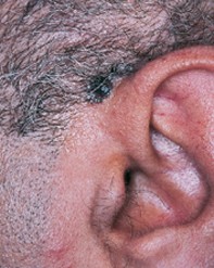
A 52-year-old man developed a progressively enlarging pigmented lesion, measuring 3.0 cm x 1.0 cm, above his left ear (Figure 1). Six years previously, a dark pigmented nodule had been removed adjacent to this site. Dermoscopy revealed an asymmetrical black lesion with an irregular border shaped vaguely like maple leaves. There were streams of pigment at the edges and focal blue–black dots. The bulk of the lesion showed structureless black pigmentation with a hazy blue–white veil (Figure 2). Excision biopsy of the lesion showed large basaloid lobules of tumour, which penetrated into the deep dermis and were associated with patchy melanin pigment (Figure 3).
Single article purchases are temporarily unavailable due to site maintenance.
If you would like to purchase an article during this time, please email us at [email protected] with the article details and we'll assist you directly. We'll also let you know when online purchasing is available again.
Thank you for your patience and understanding.