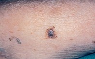
An asymmetrical mole with a blue–black centre
Skin conditions
Case presentation
A 79-year-old woman presented with a 1.4 cm diameter lesion on her left forearm. The lesion was of unknown duration but had changed in colour and increased in size. Dermoscopic examination revealed an asymmetrical lesion with an irregular, variably pigmented, broken pigment network at the periphery. The central area had an irregular, blue–black, mottled colour partially covered by a blue–grey veil. The excision specimen showed an atrophic epidermis with focal asymmetrical junctional melanocytes and large nests of melanocytes that penetrated into the deep dermis and were associated with nuclear atypia.
Single article purchases are temporarily unavailable due to site maintenance.
If you would like to purchase an article during this time, please email us at [email protected] with the article details and we'll assist you directly. We'll also let you know when online purchasing is available again.
Thank you for your patience and understanding.