Peer Reviewed
Feature Article Dermatology
Using dermoscopy to diagnose pigmented skin lesions
Recent articles on:
Skin lesions
Skin lesions
Recent articles on:
Skin cancer
Skin cancer
Abstract
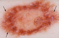
This article details a two-step method of diagnosing pigmented skin lesions using dermoscopy (surface microscopy).
Key Points
- All primary care physicians who practise in countries where melanoma causes significant mortality should learn dermoscopy.
- Step 1 differentiates melanocytic lesions from nonmelanocytic lesions, and step 2 differentiates benign melanocytic lesions from melanoma.
- To differentiate benign melanocytic lesions from melanoma (step 2), look for negative and positive features; a melanoma will have neither of the two negative features and one or more of the positive features.
- The diagnostic sensitivity of dermoscopy is not 100%. The clinical history is also important in leading to a final diagnosis.
Get full access
Buy this article
Single article purchases are temporarily unavailable due to site maintenance.
If you would like to purchase an article during this time, please email us at [email protected] with the article details and we'll assist you directly. We'll also let you know when online purchasing is available again.
Thank you for your patience and understanding.
Already a subscriber? Login here.