Beware blind spots!
Case presentations
Case 1
A 68-year-old woman presented for assessment of two skin lesions, which were diagnosed as a solar keratosis on the left cheek and a superficial basal cell carcinoma (BCC) on the presternal chest. After discussing treatment options with respect to her presenting complaints, a full skin examination was performed.
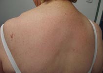
On initial inspection, the patient’s back appeared to be clear of any lesions of concern (Figure 1a). After displacing the bra-straps, a prominent vascular lesion measuring over 2 cm in maximum diameter was noted over the left upper back (Figures 1b and c). On dermoscopy, multiple cherry-red dots, globules and lacunes were observed (Figure 1d). When questioned further, the patient mentioned that the lesion was longstanding.
The provisional clinical diagnosis was angioma serpiginosum. A biopsy was not considered necessary. The patient was reassured that the lesion was benign and not requiring treatment.

