Assessing eye problems in children
Vision impairment and blindness
Eye diseases
The assessment and management of eye problems in children can be challenging. An understanding of common presentations can help guide appropriate referral.
- Examining children’s eyes can be challenging; age-appropriate tests of visual acuity and ocular alignment are required.
- A child with strabismus requires careful ophthalmic assessment to diagnose the underlying cause.
- Watery, sticky and/or red eyes are common in children; knowledge of the common causes of each symptom is helpful in guiding the assessment.
- Most cases of nasolacrimal duct obstruction resolve by 12 months of age.
- Preseptal cellulitis must be treated aggressively to prevent extension into sight-threatening orbital cellulitis.
- Amblyopia prevention and treatment remains one of the primary goals when dealing with eye conditions in children.
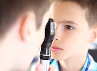
Picture credit: © Gilaxia/iStockphoto.com Models used for illustrative purposes only
Parents may present with a variety of concerns about their children’s eyes. Common presenting symptoms or signs and the most common associated conditions are listed in the Table. It is important to take a history from the parents about their specific ocular concerns as well as the child’s general health and development, prematurity and any family history of eye problems.
Paediatric ophthalmic assessment
Visual acuity
Several clues about the child’s visual abilities and possible pathology can often be gained by observing the child’s behaviour with the parent or carer in the waiting room. As with most paediatric consultations, keeping the child relaxed is made easier with the use of bright and interesting toys and by making the whole experience fun. Many different toys should be within reach to keep an infant interested in the examination.
Visual acuity testing needs to be age appropriate. Up to the age of 6 months, children should be observed to fix and follow faces or bright targets held near. Older infants should be able to fixate on distant as well as near objects, and should be able to grasp ‘hundreds and thousands’ or other small lollies.
Between the ages of 1 and 2 years, a simple test for visual acuity and visual asymmetry between the two eyes can be performed by occluding each eye separately and observing the child’s reaction or objection. Objection to occlusion is a sign of possible poor vision in the non-occluded eye. Visual acuity in preverbal children can be quantified using preferential looking tests performed by trained orthoptists.
Children older than 2 years should be able to identify pictures or match optotypes on a chart (Figures 1a and b). Older children may be able to use Snellen visual acuity charts.
Ocular alignment
Ocular alignment can be tested simply with a Hirschberg test by shining a torch at the child’s eyes and observing the light reflecting off the two corneas (Figures 2a to d). If the eyes are aligned, then the light reflection should be at the centre of each pupil. If the reflection from one eye does not correspond with the other, then strabismus is most likely present. Alternatively, a Bruckner test can be performed by looking through a direct ophthalmoscope at both red reflexes simultaneously, which should appear symmetric if the eyes are aligned. If strabismus is present then one reflex appears brighter than the other (Figure 3).
Cover testing is the observation of re-fixation eye movement of the suspected turned eye as a cover is introduced over the fixing eye. This test can be performed in infants and older children who can reliably fixate on a target. A cover test combined with the use of prisms allows the examiner to quantify the angle of the strabismus.
Cycloplegic refraction
A paediatric eye examination is not complete without a cycloplegic examination. Cycloplegia – paralysis of the ciliary muscle of the eye – is achieved by instilling cyclopentolate drops (0.5% for infants and 1.0% for older children) into both eyes and waiting at least 40 minutes. Children whose eyes dilate poorly in the clinic may be prescribed atropine 1% drops for instillation by the parents daily for three days before the next appointment. Parents should be warned about the potential side effects of cyclopentolate or atropine drops including antimuscarinic systemic side effects, which may manifest as flushing, sweating, respiratory distress and urinary retention. Atropine will blur the child’s vision for up to two weeks and children may require assistance for school activities while the drops wear off. Atropine drops should be stored in a locked cupboard out of the reach of children to prevent accidental ingestion. Excess drops should be discarded in the sink and washed away before discarding the bottle.
Cycloplegia allows measurement of the child’s focus in both eyes using a retinoscope. Lenses can be trialled with the child’s accommodation paralysed so that hypermetropia cannot be overcome by accommodation (the physiological act of changing focus from far to near). Refractive error can be hypermetropia (long-sightedness), myopia (short-sightedness) and astigmatism (oval-shaped effective defocus). Dilation of the pupils with cycloplegia also enables detailed examination of the fundus including the optic disc, macula and retina.
A significant degree of refractive error or a significant difference between the refraction of the two eyes (anisometropia) may prompt prescription of spectacles. Spectacles for children are provided by most optometrists and optical dispensers who are able to meet specific considerations including frame size, material and elastic strap.
Strabismus
Esotropia (convergent strabismus), exotropia (divergent strabismus) or vertical strabismus can be found in children. Strabismus can be intermittent or constant. All forms of strabismus are more obvious when the child is tired or unwell. Parents often bring photographs of their child who has an intermittent strabismus.
The most important step in the examination of a child with strabismus is to exclude underlying poor vision as the cause. Poor vision may be due to structural eye problems including cataracts and retinal or optic nerve pathology. Such cases of sensory strabismus are commonly associated with amblyopia. Other causes of strabismus are refractive error, usually hypermetropia, cranial nerve palsies and pseudostrabismus. The treatment of a child with any type of strabismus begins with spectacles for any refractive component and the management of secondary amblyopia if present.
Esotropia
Parents often bring their child for review after noticing him or her looking ‘cross-eyed’ or esotropic (Figure 4). Newborns and young infants can exhibit small-angle intermittent strabismus (esotropia or exotropia) until 6 months of age when the child’s vision and fixation ability improve. Children under 6 months of age with large-angle esotropia or children older than 6 months with any esotropia should be referred for assessment by an ophthalmologist.
There are several types of esotropia in children: infantile, accommodative and pseudo-esotropia.
Infantile esotropia
Infantile esotropia presents by 6 months of age, most commonly by 3 months. It is usually large angle and associated with good vision in both eyes due to cross-fixation. Children with infantile esotropia benefit from early surgical intervention to potentially restore binocular vision. Timely referral of the affected child to a paediatric ophthalmologist is recommended.
Accommodative esotropia
Accommodative esotropia usually occurs after 18 months of age when the child learns to accommodate, but can occur earlier. It is the most common form of strabismus in children. Accommodative esotropia is associated with a greater degree of hypermetropia than is age appropriate. As accommodative effort is directly linked to convergence eye movements (physiological synkinesis), the more the child accommodates, the more one eye converges. As a result, the angle of accommodative esotropia is usually larger for near than for distance.
Prolonged accommodative esotropia is not compatible with binocular vision and if untreated will lead to the development of amblyopia as the child will fixate predominantly with only one eye (usually the less hypermetropic eye). Treatment of children with accommodative esotropia is to prescribe the full hypermetropic glasses to remove the accommodative component of the strabismus (Figures 5a and b). This may require an atropine refraction to uncover the full amount of latent hypermetropia. If a residual esotropia is still present then surgical correction may be required.
Pseudo-esotropia
Pseudo-esotropia is due to the presence of broad epicanthal folds giving the illusion of esotropia when no white part of the eye is seen nasally. This is a common finding at the ophthalmologist’s practice in infants referred for strabismus (Figure 6). No treatment is required and the appearance usually improves as the child’s face grows. However, true esotropia may be present simultaneously and parents are educated to follow up to reassess the ocular alignment by means of Hirschberg and cover testing.
Exotropia
Intermittent exotropia is the most common divergent strabismus in children and has an onset between 18 months and 5 years of age. The syndrome is characterised by a divergent strabismus that is controlled by fusional mechanisms but breaks down intermittently on distance fixation or when the child is unwell or tired. Because of suppression, the child does not experience diplopia despite the large deviation. However, these children do tend to close one eye especially in bright light outdoors. This may be the presenting problem in many children and can occur before the exotropia is observed.
Treatment of intermittent exotropia is generally with observation and eye exercises. Strabismus surgery may be considered in cases of reduced binocular vision or for cosmetic reasons in older children. Exotropia may also occur after strabismus surgery for esotropia.
Acute-onset strabismus
Any child presenting with acute-onset constant strabismus requires careful clinical assessment and usually neuroimaging with MRI to exclude intracranial pathology. In addition, diplopia in children is a red flag for acquired strabismus, as above mentioned infantile and childhood strabismus conditions are not associated with diplopia. Reduced abduction and the presence of abducting nystagmus are also red flags for acquired strabismus.
Vertical strabismus
Vertical strabismus in children is most commonly caused by congenital fourth cranial nerve palsy. Vertical strabismus may not be noticeable in the primary position but can be elicited in side gaze and head tilt. The child may, therefore, have a compensatory face turn and head tilt, and this is a common aetiology of torticollis.
Poor vision
Parents may observe a young infant to have roving or searching eye movements and no definite fixation on faces. Nystagmus eye movements may also be present. The diagnostic approach to the apparently blind infant is divided into seeking bilateral anterior visual pathway pathology or cerebral pathology.
Anterior visual pathway problems are associated with nystagmus and poor pupil reactions. Common conditions may be diagnosed by taking a full history, including any hint of photophobia, nyctalopia (night blindness) or family history, and conducting a thorough examination including dilated fundoscopy and refraction.
Cerebral pathology can be divided into cortical visual impairment or delayed visual maturation. Infants with delayed visual maturation have no structural problems in visual pathway or the brain, and their vision often improves dramatically from 3 months of age, becoming age appropriate by about 6 months. In contrast, cortical visual impairment is associated with structural brain abnormalities usually in the context of other neurological or developmental problems.
An apparently blind infant should be referred in a timely manner to a paediatric ophthalmologist to assess for refractive error and structural conditions, such as bilateral cataracts, optic nerve hypoplasia, albinism and large chorioretinal colobomas. If the examination results are normal then the infant may be referred for visual electrophysiology studies to assess for occult retinal or optic nerve dysfunction. Examination by a paediatric neurologist and neuroimaging with MRI may also be required.
Amblyopia
Amblyopia is the failure to develop normal visual acuity because of abnormal early visual experience. The prevalence of amblyopia in the population is 2%.1
The causes of amblyopia are:
- pattern deprivation (‘no’ image); e.g. ptosis, cataract
- optical defocus (blurred image); e.g. refractive error including anisometropia
- strabismus (cortical suppression of one image).
Amblyopia is usually a unilateral condition but can be bilateral in cases of bilateral pattern deprivation or high refractive error. Pattern deprivation has a short critical period, around the first four months of life. Therefore, without early intervention (e.g. ptosis correction or cataract surgery) it can have a profound effect on vision in the long term. The critical period for optical defocus and strabismus is longer, up to the teenage years.
Amblyopia can develop in a child at any age and is an important consideration when treating those with various ocular conditions. Clues to the presence of amblyopia include asymmetrical visual acuity, objection to occlusion of the non-amblyopic eye and the presence of a manifest strabismus.
The treatment of children with amblyopia is with spectacles and/or part-time occlusion of the non-amblyopic eye. If a significant refractive error is found on cycloplegic refraction then spectacles are prescribed. If vision does not improve to an expected level then occlusion treatment can be performed with patching, or rarely optical blur (fogging glasses or atropine drops). Patching options include a sticky patch over the eye or over the glasses lens on one side. A sticky patch over skin is most reliable as it prevents peeking, but can cause local skin trauma and rarely infections or allergic skin reactions.
Duration of patching is based on the severity of amblyopia and age of the child. Initial improvement is usually seen over three to six months of patching therapy, which may be weaned or stopped when visual acuity is equal to the fellow eye. Children with severe amblyopia may require patching for many years. There is evidence that amblyopia due to refractive error can be treated in children up to 15 years of age and therefore treatment may be started at or extended into the early teenage years.
Conjunctivitis and watery eyes
Common causes for a child’s ‘sticky’ eyes are nasolacrimal duct obstruction and conjunctivitis including ophthalmia neonatorum. Red eyes can be due to bacterial (purulent) or viral (pink watery)conjunctivitis, foreign body and rarely herpetic disease. The child with a watery eye may have nasolacrimal duct obstruction or congenital glaucoma.
Congenital nasolacrimal duct obstruction
Congenital nasolacrimal duct obstruction occurs due to failure of canalisation of the distal nasolacrimal duct. It is the most common cause of epiphora at birth and affects 20% of newborns.2 Nasolacrimal duct obstruction is almost always associated with white eye, rather than red eyes as it would be with conjunctivitis. The differential diagnosis of epiphora in neonates also includes congenital glaucoma, which can present with larger corneal diameter and/or clouding in addition to conjunctival injection and photophobia. Neonates with these symptoms need to be referred for urgent assessment by an ophthalmologist to exclude this sight-threatening condition.
Management of children with congenital nasolacrimal duct obstruction is with regular massage to empty the lacrimal sac and observation. The natural history is spontaneous resolution in 97% of cases by 24 months of age. Antibiotics are not routinely prescribed and secondary infection, either conjunctivitis or dacryocystitis, is uncommon.
Parents are advised to perform hydrostatic massage over the lacrimal sac for 10 seconds, four times a day. The correct technique is important to the effectiveness of the manoeuvre. Parents should be shown the anatomical landmark of the anterior lacrimal crest, first on their own face, then on their child’s. Pressure is applied aiming downwards, backwards and towards the midline, using the smallest finger that can effectively apply pressure (Figure 7). The frequency of massage can be reduced if there is no mucopurulent regurgitation on the next attempt at massage. The parent’s nails may need to be trimmed and treated with a nail file to avoid skin trauma or pain to the child.
Examination under general anaesthesia combined with probe and irrigation of the lacrimal duct is recommended if nasolacrimal duct obstruction has not resolved by 12 to 18 months of age, or if it is associated with a dacryocystocoele or recurrent infection. There is a 90% cure rate with one procedure.
Intermittent recurrence of watering after spontaneous resolution or after successful probe and irrigation is usually from mucosal thickening during an upper respiratory tract infection temporarily blocking outflow of the nasolacrimal duct. This generally settles with time.
Neonatal conjunctivitis (ophthalmia neonatorum)
Ophthalmia neonatorum is conjunctivitis occurring within the first month of life (Figure 8). It typically presents with a purulent discharge within the first week and needs to be considered as a sight-threatening and potentially life-threatening condition. The most common causes of ophthalmia neonatorum are Gram-positive bacteria from the skin such as Staphylococcus epidermidis and Staphylococcus aureus; however, sexually transmitted infections from the birth canal including chlamydia, gonorrhoea and herpes simplex infection must be excluded through appropriate swabs.
Chlamydia is an intracellular infection and vigorous swab collection is needed for the highest sensitivity. An infected child should be admitted to hospital and treated with oral macrolide and parenteral cephalosporin antibiotics to cover for chlamydia and gonorrhoea until swab results are available. Topical antibiotics and eye lavage are also prescribed. The mother and her sexual contacts may also need to be treated. Chlamydial infection can be associated with pneumonitis and this needs to be excluded. Neisseria gonorrhoeae can penetrate intact cornea and lead to globe perforation within hours.
Allergic conjunctivitis
Children with allergic conjunctivitis can present with itch, irritation and red eyes. There are different types of allergic conjunctivitis. Vernal keratoconjunctivitis is characterised by giant conjunctival papillae in the upper lids (Figure 9). Seasonal or perennial allergic conjunctivitis is more common and is often associated with larger than normal conjunctival follicles. Allergic conjunctivitis is associated with refractive errors, and frequent eye rubbing can contribute to astigmatism, which can become amblyogenic.
Foreign body
Ocular surface foreign bodies may be removable using a cotton bud under topical anaesthesia with the child awake. However, after the first trial a child will often not allow subsequent attempts, therefore removal under general anaesthesia may be necessary in children under the age of 5 years. This is especially important for foreign bodies on the cornea.
Herpetic eye disease
Herpes simplex virus infection is a great mimicker and is often overlooked in children with red eyes. It can cause blepharitis (Figure 10) and/or keratoconjunctivitis and is invariably unilateral in presentation. Children with primary herpes simplex virus infection are treated with systemic antiviral agents. Herpetic keratoconjunctivitis can be recurrent and affected patients are treated with topical aciclovir. Repeated episodes can lead to corneal scarring and reduced vision.
Eyelid lumps and swelling
Preseptal cellulitis
Preseptal cellulitis is a common condition affecting infants and young children. Affected children usually present with a swollen erythematous eyelid (Figure 11). Pain and tenderness develop as the infection progresses. It is important to ensure that the child’s visual acuity, pupil reactions, eye movements and the globe itself are normal. Preseptal cellulitis is usually related to sinus disease and parents often give a history of a recent upper respiratory tract infection. Other causes include dacryocystitis, trauma causing a skin break (such as an insect bite) or rarely secondary bacterial infection in association with a chalazion.
Preseptal cellulitis in children progresses more rapidly than in adults due to the thinner orbital septum and higher incidence of sinus disease. As a result they must be treated more aggressively with regular review and a low threshold for admission to hospital. A trial of a broad-spectrum oral antibiotic such as amoxicillin with clavulanic acid is appropriate for mild or early infections in older children as long as parents are able to attend hospital urgently should the child’s condition worsen. Any child who is systemically unwell or has rapid worsening in eyelid swelling should be admitted to hospital for intravenous broad-spectrum antibiotics and assessment by an ophthalmologist.
Orbital cellulitis
Orbital cellulitis refers to an infection spreading into the orbital cavity, usually secondary to a sinus infection. Affected children are systemically unwell and may have reduced vision, double vision, limited eye movements, a relative afferent pupil defect or proptosis (Figures 12a and b). These children require urgent admission to a tertiary children’s hospital for multidisciplinary care including orbital imaging and high-dose intravenous antibiotics. Drainage of orbital collections and sinus washout may be required by specialist paediatric oculoplastic and ear, nose and throat (ENT) surgeons.
Chalazion
A chalazion is a lipogranulomatous inflammation occurring secondary to obstruction of a Meibomian gland. It usually presents as a non-tender erythematous lump near the eyelid margin (Figure 13). More than one lump may be present in one or both lids. Antibiotic treatment is not required unless secondary preseptal cellulitis occurs, which is rare. Treatment of children with chalazion is conservative with a warm compress followed by lid massage twice a day. Improvement is seen gradually over weeks to months, and more than 90% of cases resolve by one year. Surgical incision and drainage may be required for social reasons (e.g. cosmetic concerns) and larger lesions that affect vision owing to ptosis or astigmatism.
Dacryocystocoele
A dacryocystocoele is associated with 0.1% of cases of congenital nasolacrimal duct obstruction.2 It presents as a blue lump in the medial canthus at birth (Figure 14a). It contains expressible mucus and amniotic fluid (Figure 14b). Children with a dacryocystocoele require early intervention with firm compression and massage four times a day.
Dacryocystocoeles may be associated with intranasal cysts particularly when bilateral. As infants are obligate nasal breathers, if there is any concern about cyanosis particularly during feeding, a referral to an ENT specialist is required. If the dacryocystocoele has not resolved by 2 weeks of age then formal probe and irrigation of the lacrimal system is recommended.
Dacryocystitis
Dacryocystitis is an infection of the lacrimal sac, presenting with a lump at the medial canthus with overlying cellulitis (Figure 15). This may occur in up to half of cases of dacryocystocoele. The organism is usually a Streptococcus sp. in patients with acute infections, whereas chronic dacryocystitis may be due to infection with Streptococcus, Proteus or other organisms. A swab is taken for culture and the child admitted to hospital for administration of intravenous antibiotics. Incision and drainage may be required, but otherwise probe and irrigation is performed after resolution of the acute infection. Some children may require a dacryocystorhinostomy performed by an orbital surgeon.
Periocular haemangiomas
Infantile haemangiomas are often located in the periocular region and present after birth but within the first six months of life. These may be associated with orbital haemangiomas, ptosis and astigmatism. Half of these children will have some degree of amblyopia. The assessment and management of these children is multidisciplinary through ophthalmology, paediatric and vascular malformation (usually run by dermatologists) clinics. After assessment for the extent of the haemangioma and possible associated syndromes, treatment of children with orbital haemangiomas is with oral propranolol on an outpatient basis.
Other conditions
Leukocoria
Leukocoria refers to a pupillary light reflex that is white rather than the normal red colour (Figure 16). Leukocoria is assumed to be due to the potentially life-threatening cancer retinoblastoma until proven otherwise. Any child with an abnormal or asymmetric pupillary reflex noted in regular lighting or in photographs should be reviewed by an ophthalmologist.
The most common cause of an abnormal pupil reflex is pseudoleukocoria caused by an off-centre camera flash, ocular misalignment or refractive error. Pathological causes of leukocoria are persistent fetal vasculature, congenital cataract and retinoblastoma. A child with retinoblastoma requires urgent assessment and treatment.
Retinopathy of prematurity
Retinopathy of prematurity (ROP) was historically a common cause of visual impairment in premature children with intact cognitive and neurological development. Because of effective screening and treatment of high-risk premature infants in neonatal intensive care units, blindness from ROP is now uncommon in developed countries. Risk factors for ROP include low birth weight (<1250 g) and low gestational age (<31 weeks’ gestation). Of these children who are screened, 2.5% will develop sight-threatening ROP requiring treatment with laser. Premature infants (with or without ROP) are at increased risk for amblyopia, strabismus and refractive errors.
Paediatric ptosis
Ptosis in children is commonly due to congenital malformation of the levator muscle (Figure 17). Other causes include third nerve palsy, an upper eyelid or orbital mass, and Horner syndrome. Children with ptosis should be referred to an ophthalmologist for urgent assessment and treatment including management of secondary amblyopia.
Periocular trauma in children
Blunt globe trauma can result in hyphaema (blood in the anterior chamber), as well as traumatic mydriasis and retinal oedema (commotio retinae). Severe blunt trauma can lead to globe rupture. A child with hyphaema needs to be assessed by an ophthalmologist and is generally admitted to hospital for observation and limitation of
physical activity. The degree of hyphaema and intraocular pressure are monitored daily. A topical corticosteroid such as prednisolone is generally prescribed in addition to cycloplegia. Re-bleeding may occur at days three to four after the initial injury.
Facial trauma in children can result in a white-eye blowout fracture of the orbital floor, in which a greenstick mechanism of fracture results in entrapment of the inferior rectus/inferior oblique muscle or its associated connective tissue. An eye examination may be normal apart from limitation in eye elevation, which may also be associated with a vasovagal response.
Penetrating trauma can result in eyelid lacerations including damage to the lacrimal drainage apparatus. In addition, globe injury can occur with corneal or scleral perforation. The pupil may be irregular in shape with a shallow anterior chamber. A child with a penetrating trauma requires urgent referral to an ophthalmologist. A plastic shield or the end of a clean styrofoam cup should be applied over the child’s eye without an eye pad and the child kept nil-by-mouth until assessment. Intravenous antibiotics, pain relief, antiemetic drugs and tetanus toxoid are given if required. Surgical repair under general anaesthesia is needed after assessment of the full extent of the injuries.
Any child with periocular trauma needs a dilated fundus examination. The possibility of nonaccidental injury must be considered in all children and followed up as required.
Syndromes associated with ocular involvement
- Trisomy 21 is associated with blepharitis, nasolacrimal duct obstruction, refractive error, strabismus, amblyopia, epicanthal folds, congenital cataract and glaucoma.
- Neurofibromatosis type I is associated with eyelid neurofibromas, Lisch nodules of the iris, optic pathway gliomas and secondary glaucoma.
- Juvenile idiopathic arthritis may be associated with asymptomatic uveitis, which can lead to cataract and glaucoma; these children are, therefore, screened by ophthalmologists at appropriate intervals based on risk factors including sex, age of onset and antinuclear antibody positivity.
- Developmental delay is associated with refractive error, strabismus and amblyopia.
Vision screening programs
Screening programs are aimed at identifying children with poor or unequal vision between the two eyes during this critical period of development. Initiation of occlusion therapy at any stage may improve the visual acuity in the affected eye.
Screening programs vary between states and territories. In NSW, the Statewide Eyesight Preschooler Screening (StEPS) program targets 4-year-old children at preschool and in child care centres and offers them free screening. Children are offered screening in the first year of school in the ACT and in year 1 at school in Queensland. In Victoria, 3.5-year-old children are offered screening through the Maternal and Child Health Service with the Melbourne Initial Screening Test (MIST). GPs can check vision in children aged between 3.5 and 5 years as part of a general health assessment.
How well children recognise letters is variable in the 3- to 4-year-old age group. Therefore, screening test results may capture those who are borderline performers.
Vision and learning disabilities
Ophthalmologists are frequently consulted for an opinion regarding learning difficulties in children including, but not limited to, dyslexia. Sometimes an easily treatable cause can be found, such as undiagnosed refractive errors. Ocular motility disorders are less likely causes. There is a lack of scientific evidence in support of behavioural or perceptual vision therapies such as training or specially tinted spectacles.
Conclusion
Children generally do not complain about visual symptoms or difficulties and therefore rely heavily on parents, community and primary health workers in identifying clues such as strabismus or a learning difficulty. Awareness of the common childhood eye problems will help guide appropriate referral and expectations of the diagnostic pathway.
A child attending a paediatric ophthalmologist is expected to have an eye examination with the pupils dilated with a cycloplegic agent; cycloplegia lasts a day but occasionally a week. Amblyopia prevention and treatment remains one of the primary goals when dealing with eye conditions in children. MT
References
- Pai AS, Rose KA, Leone JF, et al. Amblyopia prevalence and risk factors in Australian preschool children. Ophthalmology 2012; 191: 138-144.
- MacEwen CJ, Young JD. Epiphora during the first year of life. Eye 1991; 5: 596-600.
Further reading
- Handler SM, Fierson WM, Section on Ophthalmology; Council on Children with Disabilities; American Academy of Ophthalmology; American Association for Pediatric Ophthalmology and Strabismus; American Association of Certified Orthoptists. Learning disabilities, dyslexia, and vision. Pediatrics 2011; 127: e818-e856.
- Kennedy SA, Noble J, Wong AMF. Five things to know about esotropia. CMAJ 2012; 184: 1279.
- Lyons C. Where the wild things are: when esotropia misbehaves. Am Orthopt J 2012; 62: 61-69.
- Pediatric Eye Disease Investigator Group. The clinical spectrum of early-onset esotropia: experience of the Congenital Esotropia Observational Study. Am J Ophthalmol 2002; 133: 102-108.
COMPETING INTERESTS: None.