Eye strain: focus on causes and treatment

Vision impairment and blindness
Eye diseases
Eye strain is relatively common and can have a major impact on work or study. GPs are ideally placed to assess contributing factors, exclude potentially serious conditions that have similar symptoms and facilitate specialist referral.
- Eye strain (asthenopia) is relatively common across all age groups and can lead to a range of nonspecific symptoms, including ocular fatigue, discomfort, tearing, blurred vision, diplopia and headaches.
- Patients with eye strain often present to a GP as the initial point of care.
- It is important for GPs to exclude potentially serious conditions that present with similar symptoms and require immediate referral and treatment.
- After eye strain is diagnosed, contributing causes may not be readily apparent and investigation of both ocular and general health may be required.
- GPs are well placed to guide the general care and referral pathway for patients with eye strain.
- A combined approach involving the GP, ophthalmologist and other specialists may be required.
Eye strain (asthenopia) refers to a collection of nonspecific ocular symptoms, including ocular fatigue, discomfort, tearing, blurred vision, diplopia and headaches. GPs are often the initial choice for patients seeking investigation of eye symptoms. As a wide range of conditions may present with symptoms similar to eye strain, it is important not to miss a potentially serious condition that requires immediate referral and treatment (Box 1).
The impact of eye strain varies considerably, from mild discomfort to significant disruption that may reduce efficiency or require time off study or work. Identifying the primary cause can be difficult, and a multidisciplinary approach involving the GP and eye care professionals is often required.
In this article, we outline the causes, presentation and diagnosis of eye strain. We also discuss how GPs can identify factors contributing to eye strain and formulate the most appropriate referral pathway.
Epidemiology of eye strain
Eye strain can affect all levels of the population but appears to be most frequent in the second and third decades of life, with a general decline in incidence from the fourth decade.1 Unsurprisingly, the peak incidence corresponds to a time of increased visual stress for many people, during secondary and tertiary education and initial career opportunities. In a large series of university students, over half (57%) complained of eye strain at some stage.2 Symptoms were significantly associated with computer use and influenced by psychosocial state, dietary habits and environmental conditions, suggesting that a broad approach to diagnosis and treatment is essential.
The incidence of eye strain symptoms in schoolchildren is lower but also considerable. Symptoms were reported by 20% of a sample of schoolchildren in Korea and 23% of a young Swedish population aged 6 to 16 years.3,4 Of note in the Swedish group, all but two of the children with symptoms had abnormal results on eye examination, confirming the importance of ophthalmic investigation in young people with eye strain.
Career and work environment are important considerations. A study found that eye strain is more prevalent in professional computer users who undertake long hours of continuous computer work.5 There was an additional association with female sex, which has been found elsewhere, albeit in small cohorts.6
Pre-existing eye conditions affect the incidence of eye strain.4,5 Uncorrected refractive error or extraocular muscle imbalance can increase symptoms of eye strain. Visual stress has also been reported in other conditions. For example, up to 20% of patients with dyslexia reported visual fatigue, visual perceptual distortions and headaches after episodes of repetitive near work.7
Symptoms of eye strain
Symptoms of eye strain vary considerably between patients and may include headaches, general eye discomfort or fatigue and visual distortions. Ocular symptoms particular to eye strain may include distance blur (when looking from near to far), difficulty refocusing, diplopia, increased light sensitivity, dry eye and a burning sensation.6 A strong indicator of visual stress is the perception of words ‘moving’ when reading, or tired uncomfortable eyes after prolonged near work.8
Causes of eye strain
A combination of ocular, general health and environmental factors can contribute to eye strain (Box 2).
Ocular causes
Specific ocular symptoms may suggest the possible cause. A study found that people with uncorrected refractive error were more likely to report eye strain and headaches.9 Poor or reduced corrected acuity (with glasses or contact lenses) increases the difficulty of visual tasks and can exacerbate eye strain.
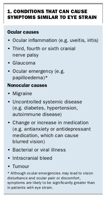
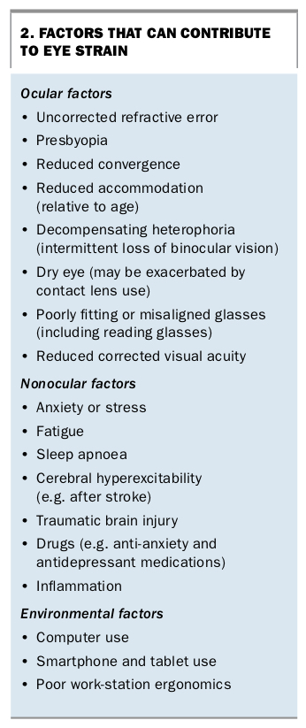
Reduced convergence (ability to maintain a single image of an object at close range) may lead to migraine, eye pain and diplopia, particularly after extended near work (Figures 1a to c).10 Poor accommodation (ability to focus at near distance) may lead to difficulty in focusing from near to far (distance blur) or general blur during reading, which patients may describe as ‘floating’ or ‘moving’ print (Figure 2).11 Accommodation is age-dependent and may also be influenced by refractive error or general health. Decompensated ocular motor function leading to intermittent or constant strabismus (turned eye) can cause diplopia, particularly in adults.
Optical devices can contribute to ocular symptoms. Extended contact lens use and poor blinking function can lead to intermittent blur and dry eye. Poorly fitting or misaligned glasses, as may occur with over-the-counter reading glasses, can increase ocular strain leading to blur or diplopia.12 Foreign body sensation, light sensitivity and fluctuations in vision, which are common in patients with clinical dry eye, can exacerbate general ocular discomfort and strain.
General health contributing factors
Nonocular causes of eye strain are relatively common. Fatigue and anxiety can exacerbate general ocular symptoms. Significant ocular fatigue and restriction of the effective field of vision have been identified in drivers with sleep apnoea compared with normal subjects, highlighting definitive practical concerns.13
Visual stress resulting from cerebral hyperexcitability occurs in a range of neurological conditions, including stroke.14 Symptoms may include headaches, glare complaints and eye strain with minimal provocation.
Traumatic brain injury can have a significant impact on the visual system leading to eye strain, reading problems, blurred vision, diplopia and vertigo.15 Early review of visual capabilities after the trauma may be key to minimising general functional problems and facilitating recovery. Postinjury medications, including anti-anxiety medications, anticonvulsants and antidepressants, have been implicated in blurred vision and eye strain, but the effects may be indistinguishable from those of the injury itself.16
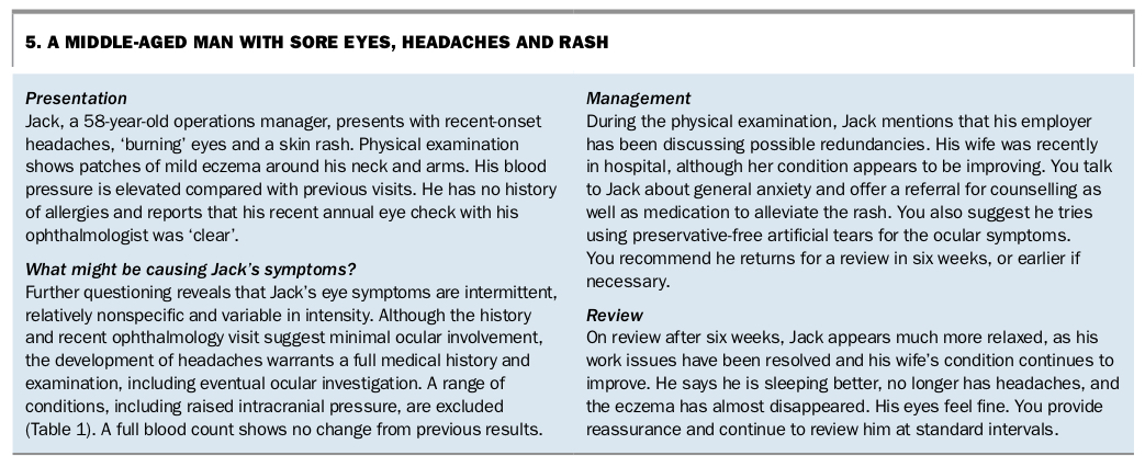
Anxiety can manifest with general symptoms. For example, it was recently suggested that anxiety about job security may lead to increasing headaches, eye strain and general concerns such as skin problems, albeit in a small subgroup of patients who are more responsive to stress conditions.17
In rare cases, eye strain has been the presenting symptom of inflammation. For example, in a retrospective review of 100 consecutive cases of allergic fungal sinusitis, 34% of patients reported ocular symptoms, including visual disturbance, epiphoria and double vision.18 In a separate case report of a patient with allergic fungal sinusitis, inflammation led to displacement of the medial rectus muscles, which induced symptoms of eye strain. These symptoms improved after surgical treatment.19 This highlights the variety of potential contributing factors to eye strain.
Environmental causes
The increasing use of near vision in both work and leisure activities may contribute to visual discomfort, although the effect varies between individuals. Computer use for longer than six hours per day was found to lead to visual symptoms in 72% of users, with a quarter describing moderate to severe complaints. Patients in the pre-presbyopia stage (about age 40 to 45 years) were likely to be more affected, because of declining accommodation ranges.6 Decreased blink rate, common among computer users, can increase dry eye symptoms leading to ocular discomfort, which may be exacerbated by airconditioning.
The type of computer monitor used may also influence discomfort. A study found that the incidence of eye pain in volunteers was greater after use of flat-screen monitors than curved-screen monitors.11 The authors proposed that the monitor type influenced the near point of accommodation. Three-dimensional (3D) and virtual reality units are increasingly used for entertainment. Although results of studies on their impact vary, the literature supports a possible role of 3D animation and movies in increased visual fatigue.20 The term ‘computer vision syndrome’ has been suggested to describe the collection of symptoms related to close work but would be synonymous with eye strain.21
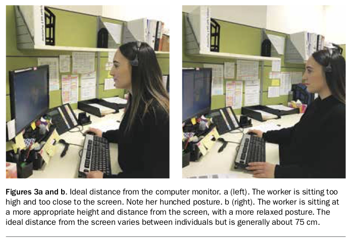
Poor ergonomics of the work station, including inappropriate intensity and direction of lighting, glare and poor positioning of monitors can manifest as both visual and musculoskeletal problems (Figures 3a and b). Ergonomics should be considered if no contributing ocular or general health conditions are identified and eye strain symptoms do not resolve with basic treatment.22
Research also highlights how the use of devices such as smartphones and tablets can contribute to visual stress. Smartphone users were found to move the viewing distance of their phones closer with prolonged use (60 minutes or more), increasing eye strain symptoms in some users.8 Furthermore, users tended to hold their devices closer when they were lying down than when sitting.8
Diagnosis
Ocular symptoms such as sore, gritty and watery eyes, ocular fatigue or discomfort, intermittent blurred vision, headaches or double vision can be caused by eye strain or a wide range of other conditions. In patients with these symptoms, it is important for GPs to identify any potentially serious conditions that require immediate referral and treatment (Box 1).
Detailed history-taking, examination and investigation are imperative. Questions for the GP to consider include:
- Does the patient also have sinus problems?
- If they have a headache, what is the cause?
- Could it be something more sinister (e.g. a brain tumour or cerebral bleed)?
- Is it uveitis, glaucoma or an ocular emergency?
- Does the patient have a systemic health problem (e.g. diabetes, an autoimmune process or uncontrolled hypertension, which can all have eye-related signs and symptoms)?
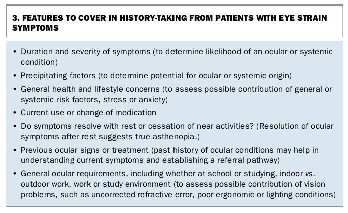
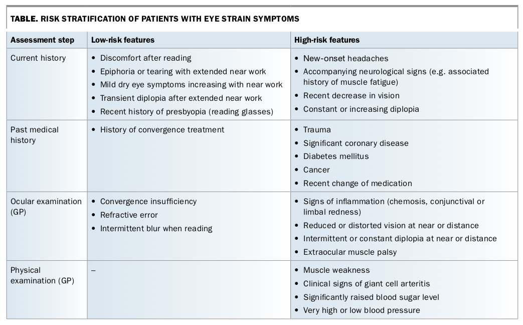
Points to cover in history-taking in a patient with eye strain symptoms are summarised in Box 3. Low- and high-risk features of the history and of both ocular and physical examination are shown in the Table.
Presentation with headache
In patients who present with headache, it is important to ask about vision and visual tasks as well as assessing for more sinister problems, as many patients do not associate headache with eye problems. It is also important to explore the history of the headaches, their duration, severity, onset and precipitating and alleviating factors, the patient’s hydration status and any history of syncope or trauma.
Physical examination of patients with headache should include:
- assessment of vital signs, especially blood pressure
- neurological examination, including fundoscopy
- a systems review.
Investigations such as a full blood count, multiple biochemistry analysis and thyroid function tests are needed to exclude more serious or potentially reversible abnormalities, such as anaemia, renal or hepatic disorders, electrolyte imbalances or thyroid dysfunction. Measurement of erythrocyte sedimentation rate (ESR) and C-reactive protein (CRP) level is important to diagnose temporal arteritis or other inflammatory conditions. If the ESR or CRP level are within the normal range but temporal arteritis is still suspected then a biopsy of the temporal artery should be arranged. If headaches are severe and persistent or pathology is suspected, then CT or MRI scanning may be indicated.
Identifying the cause of eye strain
In patients with suspected eye strain, the next step is to identify the underlying cause. Diagnosis of a single contributing factor can be difficult and may require a multidisciplinary approach. However, the GP is ideally placed to assess the contribution of individual factors, including general health and possible psycho-social concerns, as described previously (Box 3).
Visual history
The history from a patient with eye strain symptoms should cover eye use, near tasks, hobbies, screen time, and work and school ergonomics and environments. In children, it is important to ask where they sit in the classroom and to establish whether they have difficulty seeing the blackboard or their books at near distance. It should be kept in mind that as children get older their screen time usually increases. In older patients, occupation is an important consideration. Does the patient work in an office or outdoors? Screen time, airconditioning, sunlight and reflective environments are all important factors to establish.
For a GP suspecting an eye strain-related problem, it is essential to ask about the duration of the problem or frequency of episodes to help establish a referral pathway. Questioning the patient about a personal or family history of refractive error, strabismus or eye treatment is important, as decompensating binocular conditions can lead to a recurrence of ocular symptoms.
Vision assessment
Vision testing should be routine in a patient with headaches or eye strain. Visual acuity charts are accessible to most practitioners, and reduced unaided or corrected vision can be easily identified. Patients should ideally be assessed with their current prescription if they wear glasses or contact lenses, at the correct distance and using one eye at a time. Vision improvement with the aid of a pinhole occluder can suggest uncorrected refractive error, or if optical aids are used, refractive progression. Patients with reduced vision should be referred promptly for ophthalmic assessment.
Convergence can be tested by asking the patient to focus on a target such as a pen or a fixation image that is held at arm’s length and then slowly moved towards the patient’s nose (Figures 1a and b). The near point of convergence occurs when the patient can no longer direct both eyes together on the target or describes double vision (Figure 1c). A near point of 20cm or less is poor and may lead to eye strain symptoms. A more detailed ocular examination is appropriate at this point.
Referral
In patients with headache, referral for a formal eye examination is required to rule out significant ocular pathology, including glaucoma or papilloedema. Additional ocular causes, including ocular motility or refraction changes, will also be explored at this visit. Patients with reduced unaided or best corrected visual acuity should also be promptly referred for ophthalmic assessment.
Patients with true eye strain (primary symptoms of discomfort or difficulty in working at near distance) will also benefit from a full ophthalmological examination. Although referral is still recommended in these patients, the absence of neurological or visual impairment suggests follow up is not urgent.
However, if symptoms or signs suggest a general health or neurological problem then referral for specialist assessment should not be delayed.
Treatment of eye strain
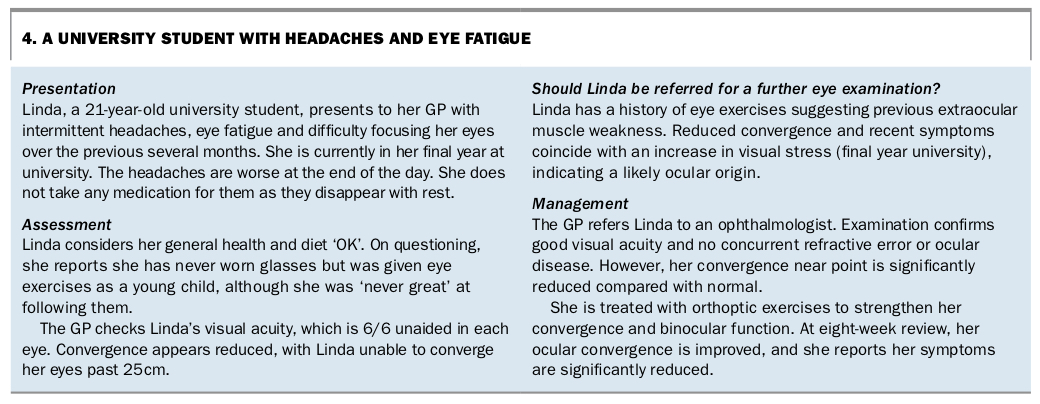
For patients who are diagnosed with eye strain, identifying the contributing factors allows treatment options to be prioritised. Case histories of two patients with eye strain and their management are shown in Box 4 and Box 5.
General measures
General health and stress-related causes should be treated according to standard practice, which may include medication or counselling. As eye strain may contribute to reduced learning and school performance, discussion with school representatives may occasionally be warranted for younger patients.
The role of supplementary vitamins in relieving eye strain symptoms is unclear. Omega-3 vitamins and bilberry extract have been found to reduce eye discomfort and ocular fatigue.23,24 This may be through improving the tear film or may reflect a previously suboptimal diet.
Ocular treatments
An uncorrected refractive error or ocular dysfunction should be treated by an ophthalmologist, optometrist or orthoptist, as appropriate. Convergence or accommodation exercises that improve the patient’s functional ocular reserves can provide benefits relatively quickly in appropriate patients.
Treatment of dry eye symptoms should be based on severity and clinical signs and titrated to the individual. This will be determined in consultation with the ophthalmologist. The use of preservative- free tears is preferred to avoid an allergic response and possible exacerbation of symptoms. Low-level dry eye symptoms generally resolve quickly with supplementary treatment. If symptoms do not improve or deteriorate then prompt referral is essential.
Ergonomic measures
Ergonomic assessment of the work or study environment may be beneficial when indicated by the patient’s history and occupation. The use of polarised computer screens, blue-blocking filters and curved monitors may help visual fatigue.25 The positioning of monitors is important. The light source should be adequate without significant glare and ideally should be directed from behind the user to avoid direct glare.
The distance and height of computer monitors can also be optimised to reduce musculoskeletal concerns. The optimal viewing distance for most people is about 75 cm from the monitor, with the upper edge of the monitor about 15cm below eye level (Figures 3a and b). However, this is a general guide and may need to be modified for additional factors such as a requirement for glasses, particularly reading glasses or multifocal lenses.26 Consideration of font size may also be appropriate.
As computer users are at greater risk of dry eyes, patients should be encouraged to blink frequently and to take short breaks from their device. Advice on viewing distance for smartphones or handheld devices may be helpful; a minimum distance of 30cm is generally recommended, although this may be modified according to patient age.
Conclusion
It is likely that most people will experience eye strain symptoms at some stage of life. These symptoms can have a considerable impact. The underlying cause may be difficult to diagnose, and patients require assessment to exclude serious underlying disease and identify possible contributing factors. GPs are well placed to guide the care and referral pathway for patients with eye strain symptoms, although assessment by an ophthalmologist or, if necessary, a neurologist must be considered when appropriate. MT
References
Single article purchases are temporarily unavailable due to site maintenance.
If you would like to purchase an article during this time, please email us at [email protected] with the article details and we'll assist you directly. We'll also let you know when online purchasing is available again.
Thank you for your patience and understanding.